Table of Contents
When it comes to diagnostic imaging, two of the most commonly used methods are CT scans (Computed Tomography) and MRIs (Magnetic Resonance Imaging). Both of these imaging techniques allow doctors to see detailed images of the inside of your body, but they work in different ways and are used for different purposes. Understanding the differences between CT scans and MRIs can help you make an informed decision about which one might be right for you, depending on your medical needs. In this blog, we’ll explore the key differences between these two imaging methods, how they work, and when each is typically used.
What is a CT Scan?
A CT scan is a type of X-ray that takes multiple images (or “slices”) of the body from different angles and combines them to create cross-sectional images. CT scans are sometimes referred to as CAT scans (Computed Axial Tomography), and they can produce detailed images of bones, soft tissues, and blood vessels. During the procedure, you’ll typically lie on a table that moves through a large, doughnut-shaped machine, which captures the images.
How CT Scans Work:
- A CT scanner uses X-rays to take detailed pictures of the inside of the body.
- These images are then combined by a computer to create a detailed 3D image of the area being studied.
- It’s often used for emergency situations, like detecting injuries after an accident, or for routine screening to assess conditions like cancers, lung diseases, or heart problems.
Benefits of CT Scans:
- Fast and efficient, especially useful in emergency settings.
- Provides highly detailed images of bones, organs, and blood vessels.
- Effective for identifying fractures, tumors, internal bleeding, and abnormalities in organs.
Limitations of CT Scans:
- Involves exposure to radiation, although the dose is generally low.
- Less effective for imaging soft tissues in comparison to MRI.
What is an MRI?
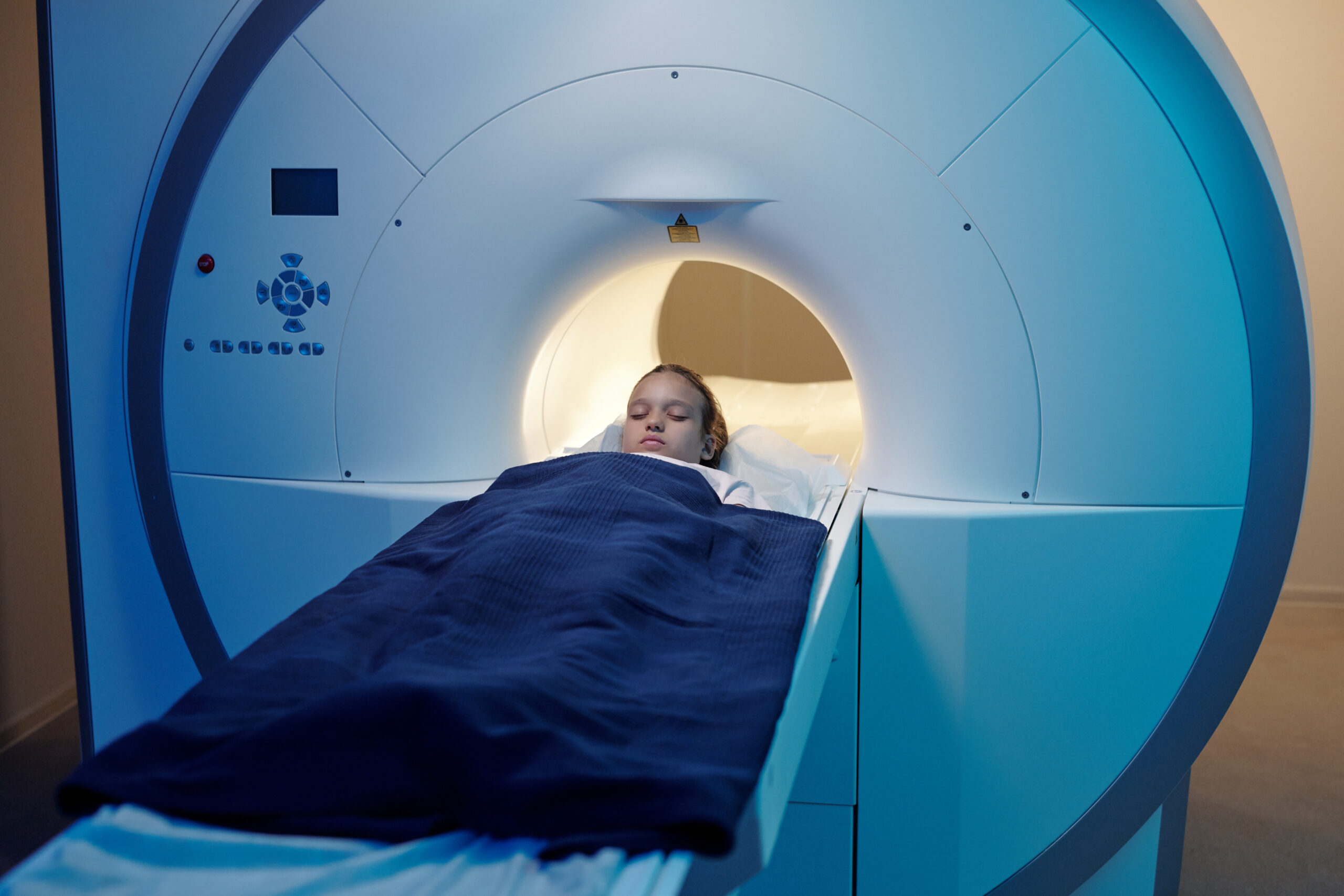
An MRI (Magnetic Resonance Imaging) is a diagnostic imaging technique that uses powerful magnets and radio waves to create detailed images of the organs and tissues inside your body. Unlike a CT scan, MRIs do not involve radiation. Instead, the magnetic field and radio waves interact with hydrogen atoms in the body to produce detailed images. MRIs are particularly useful for imaging soft tissues, such as the brain, muscles, and organs.
How MRIs Work:
- MRI machines generate strong magnetic fields that interact with the protons in the body.
- Radiofrequency pulses are sent into the body to alter the alignment of protons, and the signals emitted by these protons are used to create detailed images.
- MRIs are ideal for examining soft tissues like the brain, spinal cord, muscles, and ligaments.
Benefits of MRIs:
- No exposure to ionizing radiation, making it a safer option for repeated use.
- Excellent for imaging soft tissues, such as the brain, spinal cord, and muscles.
- Effective for detecting conditions like brain tumors, spinal injuries, joint and ligament tears, and neurological disorders.
Limitations of MRIs:
- Takes longer than CT scans, sometimes requiring 30 minutes to an hour.
- Can be uncomfortable for patients with claustrophobia due to the enclosed space of the machine.
- Not suitable for patients with certain implants, such as pacemakers or metal prosthetics, due to the strong magnetic field.
Key Differences Between CT Scans and MRIs
| Feature | CT Scan | MRI |
| Technology Used | Uses X-rays and computer processing | Uses magnetic fields and radio waves |
| Imaging Type | Best for bones, blood vessels, and organs | Best for soft tissues (brain, muscles) |
| Speed | Fast (typically 10-15 minutes) | Slower (typically 30-60 minutes) |
| Radiation Exposure | Yes, involves exposure to radiation | No, no radiation involved |
| Cost | Generally less expensive than MRI | More expensive due to longer procedure |
| Patient Comfort | Less confining, but may involve contrast dye | Can be uncomfortable, especially for those with claustrophobia |
| Use Cases | Emergency situations, bone fractures, lung conditions, cancers | Soft tissue issues, neurological disorders, joint problems |
When to Use a CT Scan
CT scans are often used when speed is essential, particularly in emergency settings. Here are some common situations where a CT scan might be recommended:
- Head injuries: CT scans can quickly identify skull fractures or bleeding in the brain.
- Lung conditions: To diagnose issues like pneumonia, lung cancer, or pulmonary embolism.
- Bone fractures: Particularly when a fracture is suspected but not visible on a standard X-ray.
- Cancer diagnosis: CT scans are effective in detecting tumors in various organs, such as the lungs, liver, or pancreas.
- Abdominal or chest conditions: Conditions like internal bleeding or gastrointestinal issues can be diagnosed with CT scans.
When to Use an MRI
MRI is preferred when soft tissue imaging is required, and is often used for chronic conditions or specialized diagnoses. Here are some common situations where an MRI might be recommended:
- Neurological disorders: MRIs are excellent for visualizing the brain, spinal cord, and nerves, making them ideal for diagnosing conditions like multiple sclerosis, brain tumors, and spinal cord injuries.
- Joint or soft tissue injuries: If you have a ligament tear, muscle injury, or joint damage, an MRI provides the detailed images needed for an accurate diagnosis.
- Cancer detection: MRI is often used to assess soft tissue tumors, such as those found in the brain, breast, and liver.
- Heart and vascular imaging: MRIs can evaluate the heart and blood vessels for conditions like heart disease and vascular abnormalities.
- Pelvic conditions: For evaluating prostate, uterine, or ovarian issues, MRI provides a clearer view than CT.

Which Is Right for You?
Choosing between a CT scan and an MRI depends on your medical condition and the part of the body being examined. Here are some factors to consider:
- Speed: If you are in an emergency situation, such as a traumatic injury, a CT scan may be the right choice because it provides quicker results.
- Radiation exposure: If you are concerned about radiation, an MRI is the better option as it does not involve any radiation.
- Soft tissue imaging: If your doctor needs to examine your soft tissues, such as the brain, muscles, or joints, an MRI is likely to provide better results.
- Bone or lung issues: For conditions involving bones, fractures, or lung problems, a CT scan would be more appropriate.
Conclusion
Both CT scan in Mumbai and MRIs are invaluable tools in the medical field, each with their specific strengths and ideal uses. A CT scan is best for fast, detailed images of bones, organs, and blood vessels, making it particularly useful in emergency situations. On the other hand, an MRI excels at imaging soft tissues and is ideal for diagnosing neurological, musculoskeletal, and soft tissue disorders.
Before deciding on which imaging technique is right for you, it’s essential to consult with your doctor. They will evaluate your symptoms, medical history, and the part of your body that needs to be imaged to determine which scan is most appropriate. Whether you need a CT scan or an MRI, both procedures are safe and effective for obtaining critical diagnostic information that will help guide your treatment and improve your health outcomes.
![]()

