Introduction
When it comes to diagnostic imaging, a CT scan in Mumbai is one of the most commonly requested tests for patients. Whether you’re dealing with unexplained pain, a serious injury, or a suspected internal condition, a CT (computed tomography) scan provides detailed images of your body’s organs, bones, and soft tissues. But with so many options for CT scans in Mumbai, how do you choose the right imaging centre that guarantees accurate, timely results in a comfortable environment?
In this guide, we’ll walk you through everything you need to know about CT scans in Mumbai—from the procedure itself to tips on selecting the best imaging centre. By the end of this article, you’ll be well-equipped to make informed decisions regarding your health and imaging needs.

What is a CT Scan?
A CT scan, also known as a CAT scan, is a non-invasive imaging procedure that uses specialized X-ray equipment and computer technology to produce cross-sectional images of the body. These images can show the structure and abnormalities of bones, organs, and soft tissues in great detail, allowing doctors to make accurate diagnoses.
CT scans are often used to detect conditions such as:
- Tumours or growths
- Injuries or fractures
- Infections or abscesses
- Internal bleeding
- Heart disease
- Strokes
- Lung diseases
The detailed images produced by a CT scan help doctors assess the severity of conditions and plan the most effective course of treatment.
Why Do You Need a CT Scan in Mumbai?
Mumbai, being one of India’s largest metropolitan cities, is home to numerous healthcare facilities and imaging centres. The demand for diagnostic imaging procedures like CT scans is rising due to increasing health awareness, advanced medical technologies, and the growing need for precise diagnostics.
Some of the key reasons why a doctor may recommend a CT scan in Mumbai include:
- Chronic Pain: If you’ve been suffering from unexplained pain, particularly in areas like the abdomen, chest, or head, a CT scan may help pinpoint the cause.
- Injury Assessment: After an accident or injury, a CT scan can help identify bone fractures, internal bleeding, or organ damage.
- Cancer Detection: CT scans are commonly used for detecting, staging, and monitoring the progression of various types of cancers.
- Heart and Lung Diseases: A CT scan can reveal problems with the heart, lungs, and blood vessels, such as blockages or infections.
- Brain Disorders: If you’re experiencing headaches, seizures, or unexplained neurological symptoms, a CT scan can provide essential information about your brain’s health.
How Does a CT Scan Work?
The process of a CT scan in Mumbai typically involves the following steps:
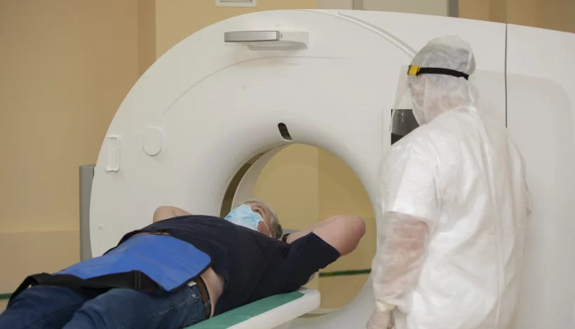
- Preparation: You may be asked to change into a hospital gown and remove any metal objects (such as jewelry or dentures) that could interfere with the scan.
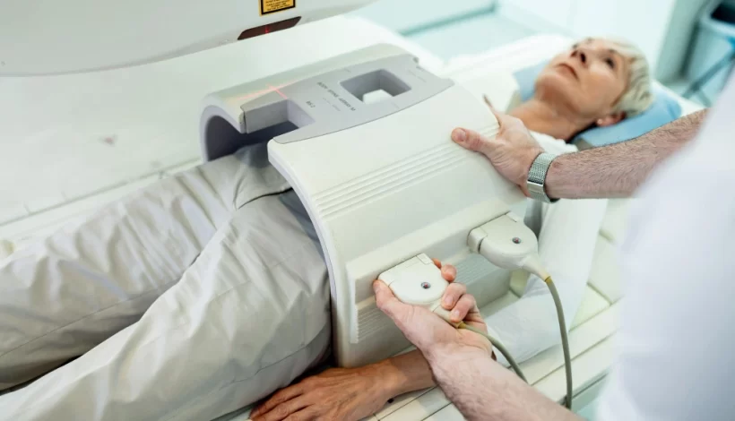
- Positioning: You will be asked to lie down on a motorized table, which will move you into the CT scanner—a large, doughnut-shaped machine.
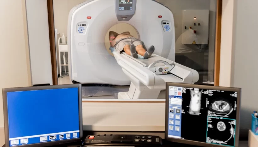
- Imaging: The scanner will rotate around your body, taking a series of X-ray images from multiple angles. These images are then processed by a computer to create detailed cross-sectional images of the body.
- Contrast Materials: In some cases, a contrast dye may be injected or ingested to enhance the images, particularly when examining blood vessels or soft tissues.
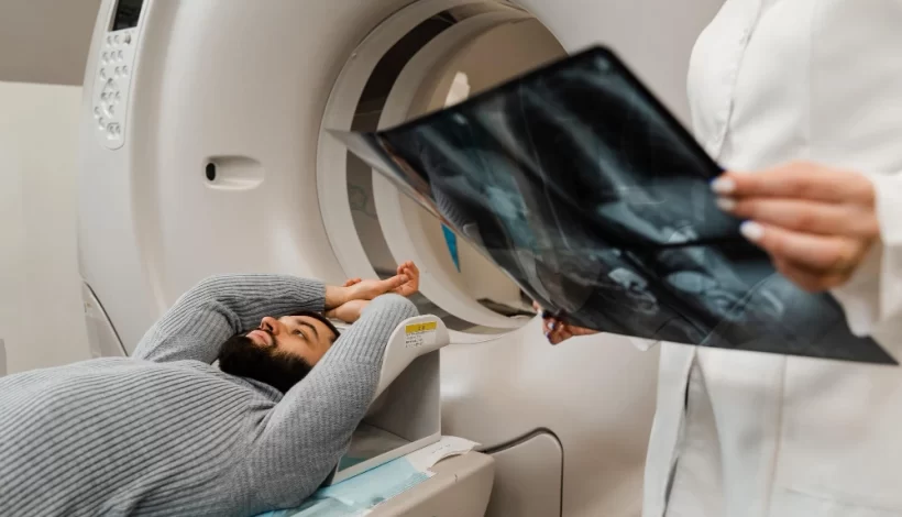
- Completion: The entire process typically takes about 10-30 minutes. Once completed, the images are sent to a radiologist for interpretation, and your doctor will discuss the results with you.
CT scans are generally painless and quick, but it’s important to remain as still as possible during the procedure to ensure clear images.
Benefits of a CT Scan
A CT scan in Mumbai offers several advantages over other diagnostic imaging methods:
1. High Precision and Detail
CT scans provide far more detailed images than regular X-rays, which allows doctors to detect conditions at earlier stages when treatment is more effective.
2. Speed
CT scans are typically fast, often taking less than 15 minutes for the procedure. This makes them ideal for emergency situations where time is of the essence.
3. Non-Invasive
Since CT scans are non-invasive, they don’t require surgery or other invasive techniques to gain a comprehensive view of your body’s internal structures.
4. Guiding Treatments
CT scans are not just used for diagnostics—they are also helpful in guiding treatments, including biopsies, surgeries, and the placement of radiation therapy for cancer patients.
5. Versatility
CT scans can examine nearly every part of the body, from the brain and heart to the abdomen and lungs. This versatility makes it one of the most commonly used imaging techniques in the medical field.
Choosing the Best Imaging Centre for a CT Scan in Mumbai
When it comes to CT scans in Mumbai, selecting the right imaging centre is crucial for ensuring accurate results and a comfortable experience. Here’s a breakdown of what to look for:
1. Accreditation and Certification
Always choose an imaging centre that is accredited by recognized medical bodies such as the National Accreditation Board for Testing and Calibration Laboratories (NABL) or the Joint Commission International (JCI). Accreditation ensures that the facility meets rigorous standards of quality, safety, and accuracy.
2. State-of-the-Art Equipment
The quality of your CT scan depends heavily on the technology used. Look for an imaging centre that uses the latest, high-resolution CT scanners. These machines deliver clearer images and reduce the need for repeat scans.
3. Expert Radiologists
Experienced radiologists who specialize in interpreting CT scan results are key to providing accurate diagnoses. Choose a centre with highly qualified radiologists with expertise in various fields, including neurology, cardiology, and oncology.
4. Patient Comfort
A good imaging centre should prioritize your comfort. Look for centres that provide clear instructions, minimize waiting times, and offer a soothing environment to reduce any anxiety you may feel.
5. Affordable Pricing and Transparency
While medical imaging can be expensive, a good centre should offer clear and transparent pricing with no hidden fees. Some centres may also offer packages or discounts for multiple scans or family members.
6. Timely Reports
Time is often of the essence when it comes to CT scans, especially if you’re dealing with a medical emergency. Choose an imaging centre that offers quick turnaround times for reports, ideally within 24 to 48 hours.
7. Positive Reviews and Reputation
Patient reviews can provide invaluable insight into the quality of service at an imaging centre. Look for centres with positive reviews that highlight professionalism, accuracy, and customer care.
Why Ace Imaging Centre is the Best Choice for a CT Scan in Mumbai
At Ace Imaging Centre, we are committed to offering the highest quality CT scans in Mumbai. Here’s why we stand out:
1. Cutting-Edge Technology
We utilize the latest CT scan machines to provide precise, high-definition images for accurate diagnosis. Our equipment ensures that your scan is performed with the utmost efficiency and safety.
2. Expert Radiologists
Our team of experienced radiologists has years of expertise in interpreting CT scans across various specialties, including neurology, cardiology, and oncology. You can trust that your results will be accurate and comprehensive.
3. Patient-Centered Care
We understand that medical procedures can be stressful, which is why we prioritize your comfort and care. From our friendly staff to our welcoming facilities, we aim to make your experience as smooth and stress-free as possible.
4. Affordable and Transparent Pricing
At Ace Imaging Centre, we offer competitive pricing for all our services, including CT scans. We believe in transparency, with no hidden charges. We are committed to providing high-quality diagnostic services without breaking the bank.
5. Fast and Reliable Reports
We know how important timely results are for your peace of mind and treatment planning. That’s why we guarantee fast report turnaround times, usually within 24-48 hours.
Conclusion
Whether you’re in need of a routine diagnostic scan or seeking answers for a serious health issue, choosing the right place for a CT scan in Mumbai is crucial for accurate results and peace of mind. At Ace Imaging Centre, we offer top-tier technology, expert radiologists, and a patient-focused approach to ensure you receive the best possible care.
Don’t compromise on your health trust Ace Imaging Centre for your next CT scan in Mumbai. Contact us today to schedule an appointment and take the first step toward better health.