The Differences Between CT Scans and MRIs: Which Is Right for You?

When it comes to diagnostic imaging, two of the most commonly used methods are CT scans (Computed Tomography) and MRIs (Magnetic Resonance Imaging). Both of these imaging techniques allow doctors to see detailed images of the inside of your body, but they work in different ways and are used for different purposes. Understanding the differences between CT scans and MRIs can help you make an informed decision about which one might be right for you, depending on your medical needs. In this blog, we’ll explore the key differences between these two imaging methods, how they work, and when each is typically used.

What is a CT Scan?

A CT scan is a type of X-ray that takes multiple images (or “slices”) of the body from different angles and combines them to create cross-sectional images. CT scans are sometimes referred to as CAT scans (Computed Axial Tomography), and they can produce detailed images of bones, soft tissues, and blood vessels. During the procedure, you’ll typically lie on a table that moves through a large, doughnut-shaped machine, which captures the images.

How CT Scans Work:

  • A CT scanner uses X-rays to take detailed pictures of the inside of the body.
  • These images are then combined by a computer to create a detailed 3D image of the area being studied.
  • It’s often used for emergency situations, like detecting injuries after an accident, or for routine screening to assess conditions like cancers, lung diseases, or heart problems.

Benefits of CT Scans:

  • Fast and efficient, especially useful in emergency settings.
  • Provides highly detailed images of bones, organs, and blood vessels.
  • Effective for identifying fractures, tumors, internal bleeding, and abnormalities in organs.

Limitations of CT Scans:

  • Involves exposure to radiation, although the dose is generally low.
  • Less effective for imaging soft tissues in comparison to MRI.

What is an MRI?

An MRI (Magnetic Resonance Imaging) is a diagnostic imaging technique that uses powerful magnets and radio waves to create detailed images of the organs and tissues inside your body. Unlike a CT scan, MRIs do not involve radiation. Instead, the magnetic field and radio waves interact with hydrogen atoms in the body to produce detailed images. MRIs are particularly useful for imaging soft tissues, such as the brain, muscles, and organs.

How MRIs Work:

  • MRI machines generate strong magnetic fields that interact with the protons in the body.
  • Radiofrequency pulses are sent into the body to alter the alignment of protons, and the signals emitted by these protons are used to create detailed images.
  • MRIs are ideal for examining soft tissues like the brain, spinal cord, muscles, and ligaments.

Benefits of MRIs:

  • No exposure to ionizing radiation, making it a safer option for repeated use.
  • Excellent for imaging soft tissues, such as the brain, spinal cord, and muscles.
  • Effective for detecting conditions like brain tumors, spinal injuries, joint and ligament tears, and neurological disorders.

Limitations of MRIs:

  • Takes longer than CT scans, sometimes requiring 30 minutes to an hour.
  • Can be uncomfortable for patients with claustrophobia due to the enclosed space of the machine.
  • Not suitable for patients with certain implants, such as pacemakers or metal prosthetics, due to the strong magnetic field.

Key Differences Between CT Scans and MRIs

FeatureCT ScanMRI
Technology UsedUses X-rays and computer processingUses magnetic fields and radio waves
Imaging TypeBest for bones, blood vessels, and organsBest for soft tissues (brain, muscles)
SpeedFast (typically 10-15 minutes)Slower (typically 30-60 minutes)
Radiation ExposureYes, involves exposure to radiationNo, no radiation involved
CostGenerally less expensive than MRIMore expensive due to longer procedure
Patient ComfortLess confining, but may involve contrast dyeCan be uncomfortable, especially for those with claustrophobia
Use CasesEmergency situations, bone fractures, lung conditions, cancersSoft tissue issues, neurological disorders, joint problems

When to Use a CT Scan

CT scans are often used when speed is essential, particularly in emergency settings. Here are some common situations where a CT scan might be recommended:

  • Head injuries: CT scans can quickly identify skull fractures or bleeding in the brain.
  • Lung conditions: To diagnose issues like pneumonia, lung cancer, or pulmonary embolism.
  • Bone fractures: Particularly when a fracture is suspected but not visible on a standard X-ray.
  • Cancer diagnosis: CT scans are effective in detecting tumors in various organs, such as the lungs, liver, or pancreas.
  • Abdominal or chest conditions: Conditions like internal bleeding or gastrointestinal issues can be diagnosed with CT scans.

When to Use an MRI

MRI is preferred when soft tissue imaging is required, and is often used for chronic conditions or specialized diagnoses. Here are some common situations where an MRI might be recommended:

  • Neurological disorders: MRIs are excellent for visualizing the brain, spinal cord, and nerves, making them ideal for diagnosing conditions like multiple sclerosis, brain tumors, and spinal cord injuries.
  • Joint or soft tissue injuries: If you have a ligament tear, muscle injury, or joint damage, an MRI provides the detailed images needed for an accurate diagnosis.
  • Cancer detection: MRI is often used to assess soft tissue tumors, such as those found in the brain, breast, and liver.
  • Heart and vascular imaging: MRIs can evaluate the heart and blood vessels for conditions like heart disease and vascular abnormalities.
  • Pelvic conditions: For evaluating prostate, uterine, or ovarian issues, MRI provides a clearer view than CT.
MRI scan in Mumbai

Which Is Right for You?

Choosing between a CT scan and an MRI depends on your medical condition and the part of the body being examined. Here are some factors to consider:

  • Speed: If you are in an emergency situation, such as a traumatic injury, a CT scan may be the right choice because it provides quicker results.
  • Radiation exposure: If you are concerned about radiation, an MRI is the better option as it does not involve any radiation.
  • Soft tissue imaging: If your doctor needs to examine your soft tissues, such as the brain, muscles, or joints, an MRI is likely to provide better results.
  • Bone or lung issues: For conditions involving bones, fractures, or lung problems, a CT scan would be more appropriate.

Conclusion

Both CT scan in Mumbai and MRIs are invaluable tools in the medical field, each with their specific strengths and ideal uses. A CT scan is best for fast, detailed images of bones, organs, and blood vessels, making it particularly useful in emergency situations. On the other hand, an MRI excels at imaging soft tissues and is ideal for diagnosing neurological, musculoskeletal, and soft tissue disorders.

Before deciding on which imaging technique is right for you, it’s essential to consult with your doctor. They will evaluate your symptoms, medical history, and the part of your body that needs to be imaged to determine which scan is most appropriate. Whether you need a CT scan or an MRI, both procedures are safe and effective for obtaining critical diagnostic information that will help guide your treatment and improve your health outcomes.

Loading

Top 5 Reasons Why a PSMA PET Scan is Recommended for Prostate Cancer Patients at Ace Imaging

Prostate cancer is one of the most common forms of cancer among men, and its detection and management are crucial to improving outcomes and quality of life. In recent years, the PSMA PET scan has emerged as a game-changer in the detection, staging, and monitoring of prostate cancer. At Ace Imaging, we are committed to providing the latest and most advanced imaging techniques to ensure accurate diagnosis and effective treatment plans for our patients. In this blog, we will explore the top 5 reasons why a PSMA PET scan is highly recommended for prostate cancer patients.

What is a PSMA PET Scan?

Before we dive into the reasons for recommending a PSMA PET scan for prostate cancer patients, let’s understand what this imaging technique entails. PSMA, or Prostate-Specific Membrane Antigen, is a protein found in high concentrations on the surface of prostate cancer cells. The PSMA PET scan is a type of positron emission tomography (PET) scan that uses a radioactive tracer designed to bind specifically to PSMA proteins. This allows doctors to accurately visualize prostate cancer cells, even in their early stages, and track their spread throughout the body.

This cutting-edge imaging technique is revolutionizing the way prostate cancer is detected, monitored, and treated.

1. Highly Accurate Detection of Prostate Cancer

The PSMA PET scan is one of the most accurate imaging tools available for detecting prostate cancer. Traditional imaging methods like CT scans and bone scans may miss small or early-stage tumors. However, PSMA PET scans provide a much higher level of sensitivity and specificity, allowing doctors at Ace Imaging to detect prostate cancer even when it is in its early stages or when it is in areas that are difficult to reach with conventional imaging.

This is particularly beneficial for:

  • Detecting Recurrence: The PSMA PET scan can help identify recurrence of prostate cancer after treatment, even if traditional methods do not show any signs.
  • Localizing Cancer Spread: It can detect the spread of cancer to lymph nodes or other distant parts of the body, which can influence treatment decisions.

With PSMA PET scans, doctors can make a more informed diagnosis and tailor treatment plans more effectively, improving the overall prognosis for prostate cancer patients.

2. Enhanced Staging and Treatment Planning

Accurate staging of prostate cancer is crucial for determining the most appropriate treatment plan. The PSMA PET scan helps determine the extent of cancer spread in a way that is far more detailed and precise compared to conventional imaging techniques. By identifying small metastases in lymph nodes, bones, or other organs, the PSMA PET scan provides valuable information that helps oncologists determine whether the cancer is localized or has spread.

At Ace Imaging, we use PSMA PET scans to assess:

  • Primary Tumor Location: Identifying the exact location of the prostate tumor to help plan surgical removal or radiation therapy.
  • Lymph Node Involvement: Detecting cancer spread to nearby lymph nodes, which may affect treatment decisions and the need for additional therapies like chemotherapy or targeted therapies.
  • Distant Metastasis: Identifying the presence of cancer cells in distant organs like the bones, liver, or lungs, which helps assess the stage and decide whether systemic treatments are necessary.

By providing a more accurate picture of the cancer’s stage, the PSMA PET scan helps doctors at Ace Imaging recommend a personalized, comprehensive treatment approach that can increase the chances of successful outcomes.

3. Non-Invasive and Safe Procedure

One of the most significant advantages of the PSMA PET scan is that it is a non-invasive imaging technique. Patients do not need to undergo painful or invasive procedures, such as biopsies, to obtain critical information about the extent of their prostate cancer. Additionally, the PSMA PET scan uses a low dose of radioactive material that is safe for patients.

The procedure involves a small injection of a radiotracer, which binds to PSMA proteins on prostate cancer cells. The patient then lies on a table while a PET scanner captures detailed images of the body. The entire process typically takes 30 to 45 minutes and requires minimal preparation. The non-invasive nature of the procedure makes it a highly preferred option for prostate cancer patients, especially those who may already be dealing with the stress and discomfort of cancer treatment.

Pro Tip: At Ace Imaging, we ensure that all our patients are made comfortable throughout the process, and our team of experts closely monitors every step of the procedure.

4. Monitoring Treatment Response and Early Detection of Relapse

One of the biggest challenges in prostate cancer management is monitoring the effectiveness of treatments and detecting early relapses. The PSMA PET scan plays a crucial role in tracking how well the cancer is responding to treatment, whether it’s surgery, radiation therapy, or hormone therapy.

As prostate cancer responds to treatment, the PSMA PET scan can show whether the tumor is shrinking or whether new areas of metastasis are developing. This allows for timely adjustments to the treatment plan. In cases where prostate cancer is hormonally resistant or has become refractory to therapy, the PSMA PET scan can identify areas of recurrence before they show up on traditional imaging scans.

By catching cancer relapse early, doctors at Ace Imaging can implement targeted treatments sooner, improving the likelihood of a positive outcome. In some cases, this could mean offering more aggressive therapies or switching to different treatment options to control the disease.

5. Personalized Treatment Decisions and Better Patient Outcomes

With its high level of accuracy, the PSMA PET scan enables a more personalized treatment approach for prostate cancer patients. Armed with detailed information about the tumor’s exact location, spread, and response to treatments, doctors can make more informed decisions about how to manage the disease.

A more personalized approach to treatment allows patients to:

  • Avoid Over-Treatment: By accurately identifying the spread of cancer, doctors can avoid over-treating non-aggressive forms of prostate cancer, preserving patients’ quality of life.
  • Tailor Therapy Plans: Whether it’s surgery, radiation therapy, or systemic treatments like chemotherapy, doctors can customize the treatment plan based on the specific needs of the patient.
  • Improve Outcomes: By using the PSMA PET scan to monitor the progression of the disease and adapt treatment as needed, patients are more likely to achieve better outcomes and longer survival rates.

Conclusion

Prostate cancer can be a challenging diagnosis, but with advancements in imaging technology like the PSMA PET scan, patients in Thane and beyond now have access to cutting-edge diagnostic tools that can greatly improve the accuracy of their diagnosis and the effectiveness of their treatment. At Ace Imaging, we are proud to offer the PSMA PET scan in Mumbai as part of our comprehensive prostate cancer care services.

Whether you’re seeking a diagnosis, exploring treatment options, or monitoring the effectiveness of your current treatment, the PSMA PET scan provides crucial information that can guide your healthcare team in making the best decisions for your health.

Loading

DOTA scan in Mumbai

DOTA Scan in Mumbai: Benefits, Process, and When It’s Needed

In the realm of advanced medical imaging, DOTA scan in Mumbai has emerged as a pivotal tool for diagnosing and managing various health conditions. Conducted at leading facilities such as Ace Imaging, this procedure provides critical insights into the body’s functional processes. In this blog, we will delve into the benefits, the process, and the specific circumstances under which a DOTA scan is recommended.

What is a DOTA Scan?

A DOTA (Diethylene Triamine Pentaacetic Acid) scan, often referred to as a somatostatin receptor scintigraphy, is a specialized imaging technique that utilizes a radioactive tracer to visualize specific cellular functions in the body. This scan is particularly effective in identifying neuroendocrine tumors and other conditions related to hormone-secreting cells.

Benefits of DOTA Scan

1. Early Detection of Diseases

One of the primary advantages of a DOTA scan is its ability to detect diseases at an early stage. This is crucial for conditions like neuroendocrine tumors, where timely intervention can significantly improve patient outcomes.

2. Non-Invasive Procedure

The DOTA scan is a non-invasive imaging technique, meaning it does not require any surgical procedures. Patients can undergo the scan comfortably without the need for anesthesia or extensive preparation.

3. Comprehensive Imaging

DOTA scans provide comprehensive imaging that helps in visualizing not just tumors but also the functionality of organs and tissues. This comprehensive view is essential for accurate diagnosis and treatment planning.

4. Minimal Side Effects

The procedure has minimal side effects compared to other diagnostic tests. Most patients can return to their normal activities shortly after the scan, making it a convenient option for many.

The Process of a DOTA Scan

Preparation

Before undergoing a DOTA scan, patients may be advised to follow certain preparatory steps, which could include:

  • Fasting: Patients are often required to fast for a few hours before the scan.
  • Medications: Inform your doctor about any medications you are currently taking, as some may need to be paused before the scan.

Procedure

The DOTA scan involves the following steps:

  1. Injection of Tracer: A radioactive tracer is injected into a vein, typically in the arm. This tracer binds to the somatostatin receptors present in certain tumors and tissues.
  2. Waiting Period: After the injection, there is usually a waiting period of about 1 to 4 hours to allow the tracer to circulate and accumulate in the target areas.
  3. Imaging: The patient will then be positioned in front of a gamma camera that detects the radiation emitted by the tracer. Multiple images will be taken from various angles to create detailed pictures of the body.
  4. Duration: The entire process, including the waiting time, can take several hours, but the actual imaging usually lasts about 30 minutes.

When is a DOTA Scan Needed?

A DOTA scan is typically recommended in the following situations:

  • Suspicion of Neuroendocrine Tumors: If a doctor suspects a neuroendocrine tumor based on symptoms or initial tests, a DOTA scan can confirm the diagnosis.
  • Monitoring Treatment Response: Patients undergoing treatment for neuroendocrine tumors may need periodic DOTA scans to assess how well the treatment is working.
  • Evaluating Symptoms: Unexplained hormonal symptoms may lead physicians to recommend a DOTA scan to check for potential underlying causes.

Conclusion

If you’re seeking a DOTA scan in Mumbai, Ace Imaging offers state-of-the-art facilities and expert medical staff to ensure a smooth experience. With its ability to provide vital information about neuroendocrine tumors and other conditions, the DOTA scan is an invaluable tool in modern medical diagnostics.

For more information about the DOTA scan process, benefits, or to schedule an appointment, feel free to contact Ace Imaging today!

Loading

MRI Scan in Mumbai

MRI Scan in Mumbai: Understanding the Process, Benefits, and Cost

What is an MRI Scan?

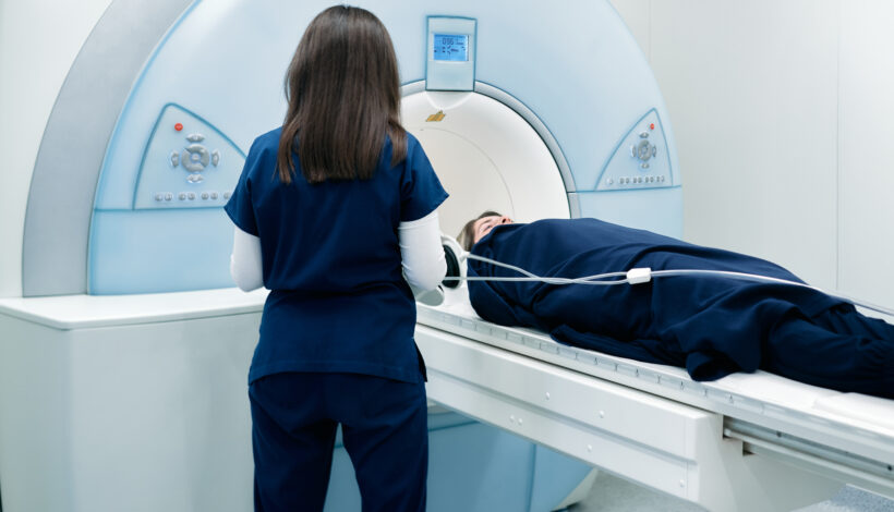
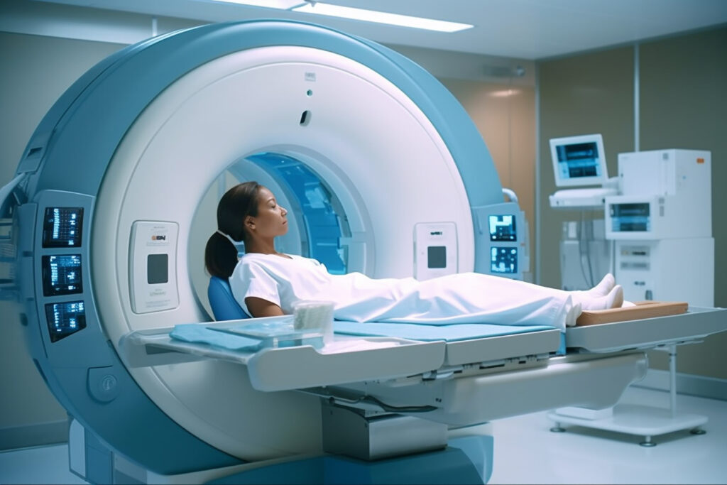
Magnetic Resonance Imaging (MRI) is a non-invasive imaging technique that uses powerful magnets and radio waves to create detailed images of the body’s internal structures. Unlike X-rays or CT scans, MRI scans do not involve radiation, making them safer for frequent use.

Why Choose an MRI Scan in Mumbai?

Mumbai is home to world-class medical facilities, including Ace Imaging, which is renowned for its cutting-edge technology and experienced radiologists. Opting for an MRI Scan in Mumbai ensures access to the latest diagnostic tools, accurate imaging, and reliable medical expertise.

Benefits of Getting an MRI Scan

  1. Detailed Imaging: MRI scans provide high-resolution images, which are ideal for detecting abnormalities in soft tissues, organs, and the nervous system.
  2. Non-Invasive and Safe: The procedure is non-invasive and does not use harmful radiation.
  3. Wide Range of Applications: MRI scans are versatile and used to diagnose various conditions, including brain injuries, spinal disorders, joint issues, and abdominal problems.
  4. Customized Scans: MRI technology can be adapted to focus on specific body areas, allowing for targeted diagnosis.

How is an MRI Scan Performed?

Getting an MRI Scan in Mumbai typically follows these steps:

  1. Preparation: Patients may be asked to change into a hospital gown and remove any metal objects to avoid interference.
  2. Positioning: The patient lies on a motorized table that slides into the MRI machine.
  3. The Scanning Process: The machine produces loud thumping or tapping noises, and patients may wear ear protection. The scan can take anywhere from 15 minutes to over an hour, depending on the body part being examined.
  4. Imaging Results: Once the scan is complete, radiologists at Ace Imaging review the images and create a detailed report for your doctor.

Cost of an MRI Scan in Mumbai

The cost of an MRI Scan in Mumbai varies depending on several factors, including the type of scan, the facility, and any additional procedures involved. At Ace Imaging, patients benefit from transparent pricing and high-quality service. Generally, MRI scan prices range from INR 5,000 to INR 15,000 or more, depending on complexity.

Factors Affecting MRI Scan Costs

  • Type of MRI: Advanced scans, such as contrast-enhanced MRIs, are priced higher.
  • Body Part: Scans for specific areas like the brain or spine might differ in cost.
  • Technology and Equipment: Facilities with state-of-the-art machines may charge more for their services.
  • Expertise of the Radiologist: Experienced radiologists who provide detailed and accurate interpretations may contribute to higher costs.

Why Choose Ace Imaging for Your MRI Scan?

Ace Imaging stands out for several reasons:

  • Advanced MRI Technology: Cutting-edge equipment ensures high-quality images.
  • Experienced Professionals: Skilled radiologists and technicians with extensive experience in diagnostic imaging.
  • Patient-Centric Care: The facility prioritizes comfort and efficient service.

FAQs About MRI Scans in Mumbai

Q1: Is an MRI scan painful?
No, MRI scans are painless. However, some patients may experience discomfort from staying still for an extended period.

Q2: Are there any risks associated with an MRI?
MRI scans are generally safe. The primary concern is for patients with certain implants, such as pacemakers, as the magnetic field may interfere with them.

Q3: How should I prepare for my MRI scan at Ace Imaging?
You may be instructed to avoid food and drink before your scan, especially if a contrast dye is used. Always inform your doctor and the radiologist of any metal implants or medical conditions.

Conclusion

Choosing an MRI Scan in Mumbai at Ace Imaging ensures a seamless diagnostic experience with expert care and cutting-edge technology. Understanding the process, benefits, and costs can help you make an informed decision about your health.

Loading