Bone pain after prostate cancer treatment can be worrying. Many patients assume it is due to ageing, weakness, or previous therapies. However, in some cases, persistent or worsening bone pain may indicate that the cancer has spread to the bones.
If you or a loved one is experiencing bone pain after prostate cancer, early evaluation is crucial. One of the most advanced diagnostic tools available today is the PSMA PET scan, which helps detect even small areas of cancer spread.
Let’s understand what this means and when you should consider advanced imaging.
Bone Pain After Prostate Cancer: When Should You Be Concerned?
After successful treatment, many prostate cancer patients remain cancer-free for years. However, in certain cases, the cancer may return (recurrence) or spread (metastasis).
The bones are one of the most common sites where prostate cancer spreads.
Bone pain that may require attention includes:
- Persistent deep aching pain
- Pain that worsens at night
- Pain in the spine, hips, ribs, or pelvis
- Pain not relieved by usual medication
While not every bone pain indicates cancer spread, it should never be ignored—especially if PSA levels are rising.
Why Does Prostate Cancer Spread to Bones?
Prostate cancer cells can travel through the bloodstream or lymphatic system. Bones provide a favorable environment for these cancer cells to grow.
Common areas affected include:
- Spine
- Pelvis
- Ribs
- Femur (thigh bone)
Early detection is essential because bone metastasis can lead to:
Spinal cord compression
Fractures
Severe pain
Reduced mobility
2. Disclose Any Medical Implants or Devices
If you have any of the following, it’s vital to inform the MRI staff:
- Pacemakers or cardiac defibrillators
- Cochlear (ear) implants
- Neurostimulators or spinal cord stimulators
- Insulin or drug infusion pumps
- Prosthetic joints or metal rods
- Vascular stents, aneurysm clips, or coils
- Programmable shunts
- Metallic dental implants or braces
- Intrauterine devices (IUDs)
- Surgical clips or staples
- Cosmetic implants or metal-infused tattoos
- Metal shrapnel or bullet fragments
Many modern implants are MRI-safe under certain conditions. However, the radiology team must verify the safety guidelines for each specific device before proceeding.
When Should You Be Concerned?
You should consult your doctor if:
- Your PSA levels are rising after treatment
- Bone pain persists for more than 2–3 weeks
- You experience sudden severe back pain
- There is weakness or numbness in legs
At this stage, advanced imaging like a PSMA PET scan becomes extremely important.
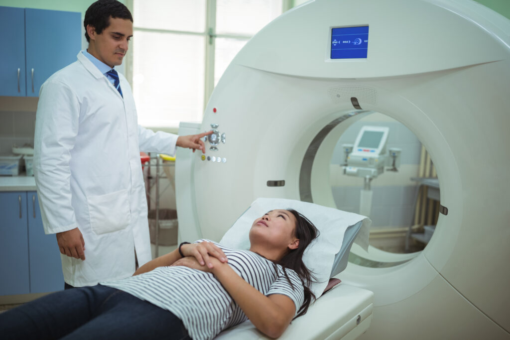
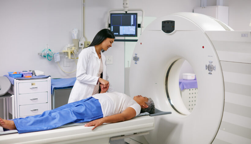
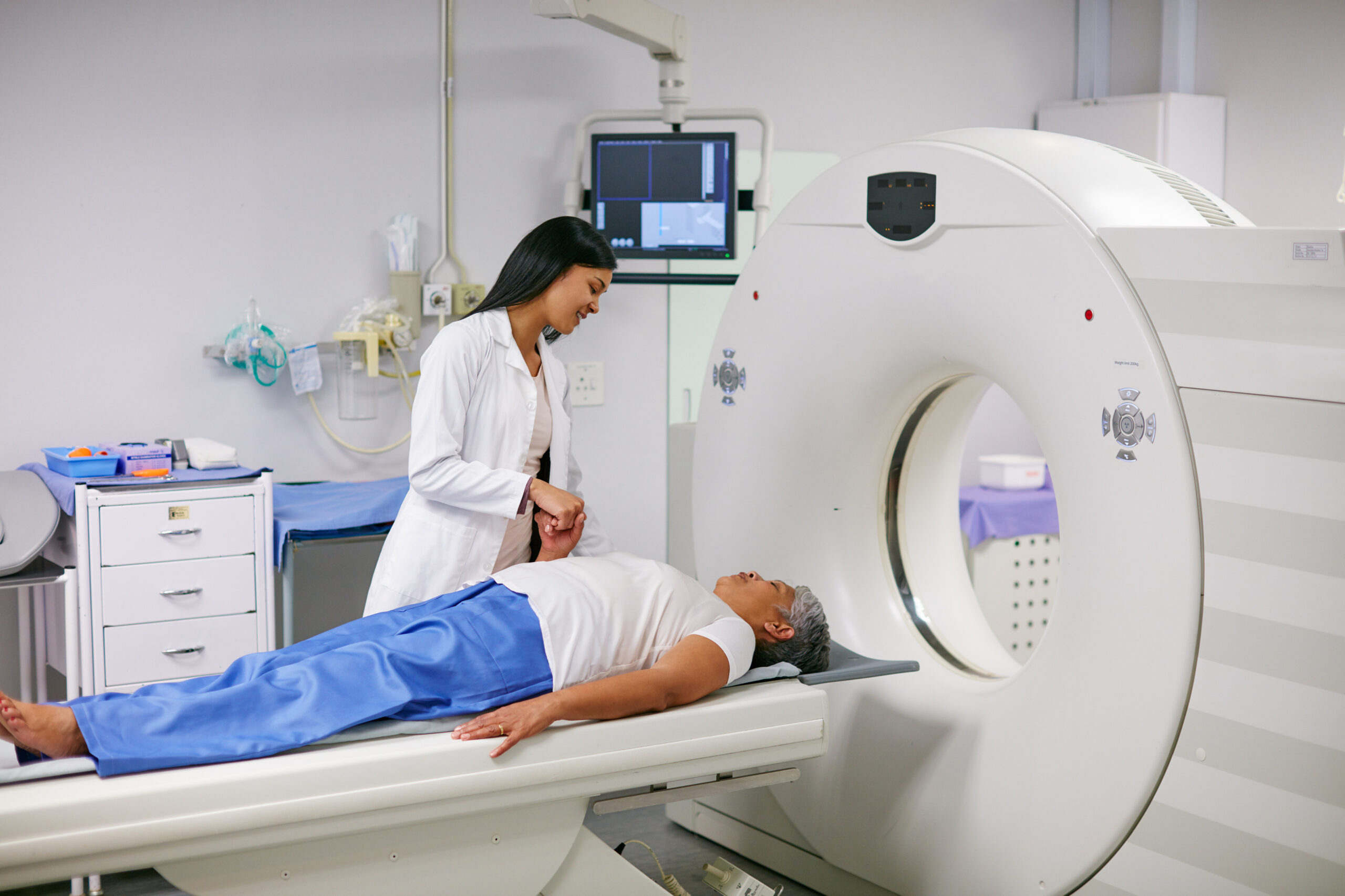
What Is a PSMA PET Scan?
A PSMA PET scan is a highly sensitive imaging test used specifically for detecting prostate cancer cells.
PSMA (Prostate-Specific Membrane Antigen) is a protein found in high amounts on prostate cancer cells. During the scan:
- A small radioactive tracer targeting PSMA is injected.
- The tracer attaches to prostate cancer cells.
- A PET-CT scanner detects even tiny cancer deposits anywhere in the body.
This makes it far more accurate than conventional bone scans or CT scanshave a slightly increased risk of rare complications related to contrast, so safety measures are important.
Can a PSMA PET Scan Detect Bone Metastasis Early?
Yes and this is its biggest advantage.
A PSMA PET scan can:
- Detect very small bone metastases
- Identify spread even when PSA levels are low
- Detect recurrence earlier than traditional imaging
- Provide whole-body evaluation in a single scan
This helps doctors:
- Plan targeted treatment
- Start therapy early
- Avoid unnecessary treatments
- Improve overall survival outcomes
Benefits of Early Detection of Cancer Spread
Detecting bone metastasis early can:
- Reduce complications
- Prevent fractures
- Improve pain management
- Help choose appropriate hormonal or targeted therapy
- Improve quality of life
The earlier the spread is detected, the better the treatment planning.
Why Choose Ace Imaging for PSMA PET Scan?
When evaluating bone pain after prostate cancer, precision matters.
Ace Imaging offers:
- Advanced PSMA PET-CT technology
- High-resolution whole-body imaging
- Experienced nuclear medicine specialists
- Accurate and timely reporting
- Patient-friendly and safe scanning protocols
Early diagnosis can make a significant difference in treatment outcomes.
Frequently Asked Questions (FAQs)
No. Bone pain can have many causes. However, in prostate cancer patients, persistent or unexplained pain should be evaluated.
It can be recommended even at low PSA levels if recurrence is suspected. Your doctor will decide based on clinical findings.
Yes. PSMA PET scans are more sensitive and detect smaller lesions compared to traditional bone scans.
Yes. It uses a small amount of radioactive tracer and is considered safe when performed under medical supervision.
The entire procedure usually takes 2–3 hours including preparation time.
Conclusion
Bone pain after prostate cancer should never be ignored. While it may be harmless, it could also indicate cancer recurrence or spread to the bones.
A PSMA PET scan provides one of the most accurate methods to detect prostate cancer spread early. Timely imaging allows doctors to start the right treatment at the right time.
If you are experiencing persistent bone pain after prostate cancer treatment, consult your doctor about advanced imaging evaluation.
Contact Information
Name: Ace Imaging
Address: 27A-B, Bezzola Complex, Next To Vijay Sales, V.N. Purav Marg, Opp. Suman Nagar, Chembur East, Mumbai-400071, Maharashtra. India
Phone:
CT – MRI Contact No
+91 22 3521 0500
+91 22 3511 2364
+91 85912 28199
Pet CT Scan
+91 22 3100 8978
+91 22 3522 2859
+91 22 3517 5845
+91 81693 60334
Website: https://aceimaging.in