In continuation with last week’s brochure on covid imaging with respiratory signs and symptoms, x-ray and HRCT chest remain the mainstay. However it is worthwhile to remember that the disease can present with multisystem involvement and atypical manifestations.
Neuro imaging
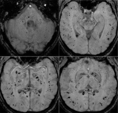
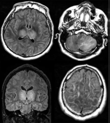
Neuroimaging reveals the following patterns: Signal abnormalities located in the medial temporal lobe / Non-confluent multifocal white matter hyperintense lesions on FLAIR and DWI, with variable enhancement, with hemorrhagic lesions / Extensive and isolated white matter microhemorrhages.

Neuroimaging figure: A

Neuroimaging figure: B
Gastrointestinal imaging
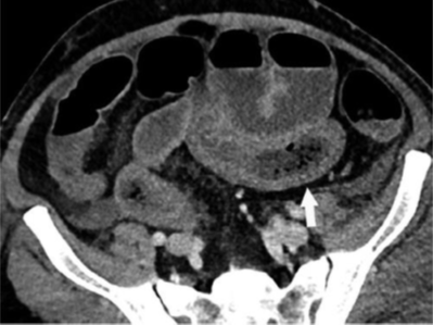
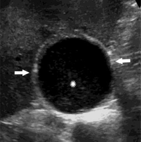
Bowel abnormalities, including ischemia, and cholestasis were findings on abdominal imaging of patients with COVID-19.

Gastrointestinal figure: A

