UNLOCK CLARITY WITH CUTTING-EDGE 1.5 TESLA MRI SCAN IN MUMBAI
“Medical Imaging is a field of rapid technological advances, and MRI has progressed from provider of gross anatomical details to a modality that enables micro-structural and physiological analysis. As compared to the 0.3 T units, the 1.5 T magnet has 5 times higher magnetic field providing higher signal-to-noise ratio, which results in better spatial and temporal image resolution. Moreover, it allows us to perform many specialized and advanced applications which are not possible on the low strength systems. Together these factors provide improved clarity and detail that will empower you to see things as you have never seen before. Combine this with our well-trained team of radiologists (having rich experience of working on advanced 1.5 T MRI systems) and you have a terrific combination of MAN and MACHINE which will provide you definitive diagnosis even in the most challenging cases. If you’re looking for an MRI scan in Mumbai, trust our expertise for accurate results. Find out about MRI scan in Mumbai cost.”
THE MACHINE
Our fully loaded 1.5 Tesla GE High Definition Signa MRI system, serving MRI scan in Mumbai, has all the latest applications for invaluable patient diagnostic evaluations.
Experience exceptional MRI scan in Mumbai with unrivaled image clarity, even in challenging cases. Trust us for precise diagnostics.
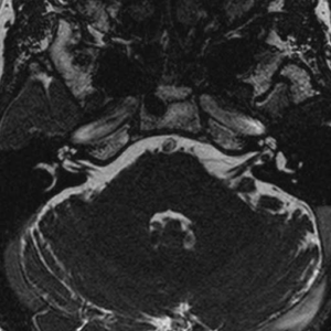
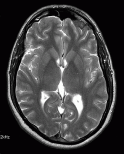
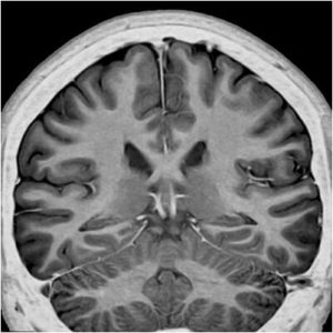
The high gradient strengths of our machine ensure great image quality each time. Moreover, the advanced “Propeller” software corrects movement artefacts and provides good image quality even in uncooperative subjects.
State of the art Neuro Applications:
Neuroperfusion
– it is now accepted worldwide as a valuable tool for characterising focal brain lesions and is especially useful in differentiating non tumorous processes from tumors ( for eg. TB granulomas from metastases and tumefactive demyelination from glioma).
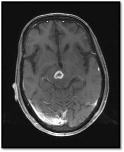
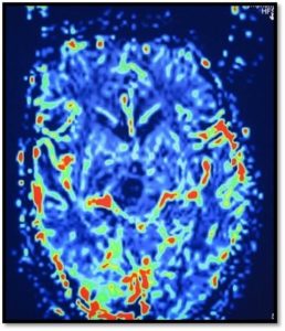
Ring enhancing lesion in right cerebral peduncle showing low perfusion (blue) on CBV map suggestive of infective granuloma (tuberculoma)
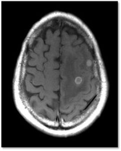
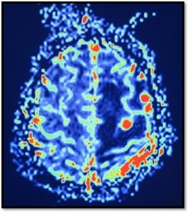
Ring enhancing lesion in left cerebral hemisphere showing high perfusion (red) on CBV map suggestive of neoplastic process (metastases)
Single and Multivoxel spectroscopy
This provides a chemical metabolite mapping of the brain lesion thus aiding its characterisation. Multivoxel spectroscopy allows evaluation of smaller lesions upto 10 mm in size and provides colour coded metabolite maps which help in planning biopsy from cell rich areas of the lesion.
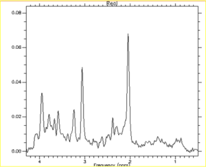
Spectroscopy graph showing normal brain metabolites.
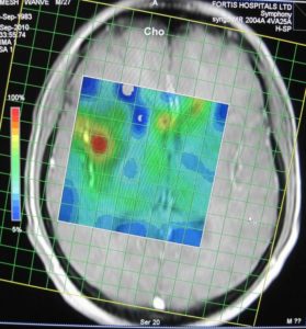
Metabolite colour map of multivoxel spectroscopy showing the high choline focus (red) which is the ideal site for biopsy
Functional MRI
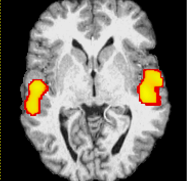
BOLD imaging allows the mapping and precise localisation of the motor strip. This is used for presurgical planning to prevent damage to the eloquent motor strip and thus reduce postoperative morbidity.
High Quality Diffusion Imaging
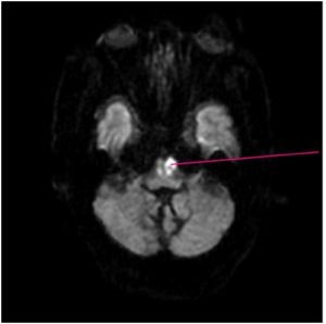
This is the most sensitive method to detect acute infarcts within minutes of the vascular event. The high resolution images can even detect the tiniest of acute lacunar infarcts. Thus now no infarct is too early or too small to be missed.
Diffusion Tensor Imaging with Fibretracking
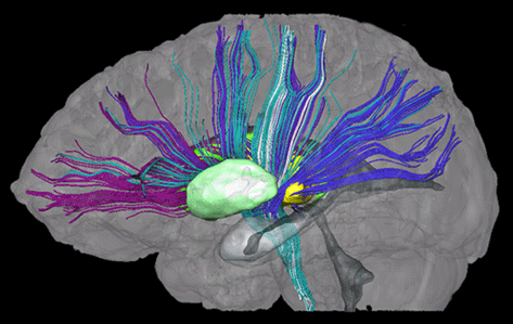
This can map the white matter tracts and help in pre-operative planning to prevent damage to these vital fibre – bundles during surgery.
CSF Flowmetry
Assesses the CSF flow dynamics and is used for diagnosis and management of Normal pressure hydrocephalus and to assess patency of surgical drainage procedures like third ventriculostomy.
MSK and Spine
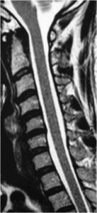
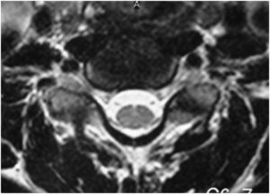
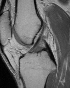
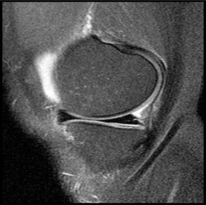
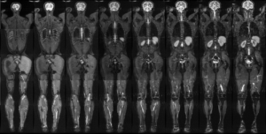
The high resolution images clearly depict the ligaments and the articular cartilage. Superior coverage allows visualisation of the entire spine in a single image. We also perform MR arthrography (especially shoulder) which provides better evaluation of the internal structures. Fast scan sequences allow the performance of MR DSA (TRICKS)which assesses the vascularity of soft tissue lesions and vascular malformations as the contrast washes in and out of the lesion.
Vascular studies
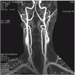
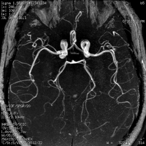
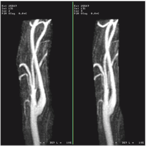
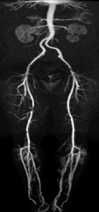
The high field and gradient strengths provide high-resolution head and neck angiographic images without the use of contrast. Fast scan times also enable contrast-enhanced angiographies in special head and neck cases and aortic, renal, and extremity angiographies. This is invaluable for patients with coexisting renal dysfunction, contraindicating iodinated contrast (used in CT and catheter angio). MR contrast (gadolinium) is less nephrotoxic, making it safer. For quality MRI scans in Mumbai, inquire about our services and MRI scan Mumbai cost.
Dynamic Pelvic Floor MRI and MR Defecography
We are probably the only center to offer this specialised study. Dynamic sequences capture the intricate pelvic floor anatomy and physiology while the patient strains and defecates. Analysis of these cine images allows precise diagnosis in patients with pelvic floor dysfunction and obstructed defecation ensuring appropriate management decisions.
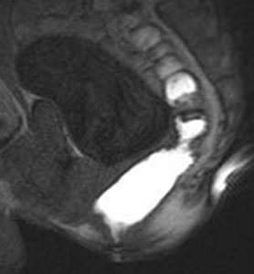
Abdominal Imaging
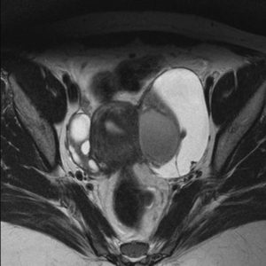
Experience top-notch abdominal imaging services at our center in Mumbai. Our advanced system ensures high-quality MRCP and MR urography images, with fast scan times for multiphasic contrast liver studies—the gold standard for liver lesion detection. Additionally, our high-resolution pelvic MRI surpasses CT for assessing pelvic disorders, including endometriosis, uterine, and adnexal lesions, as well as prostate evaluation. Explore MRI scan in Mumbai with affordable cost options.
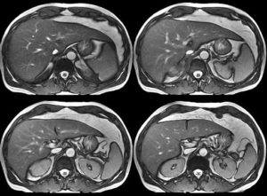
T2w liver MRI
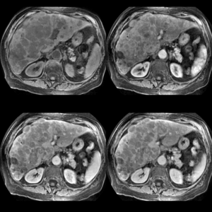
Multiphasic dynamic liver contrast MRI
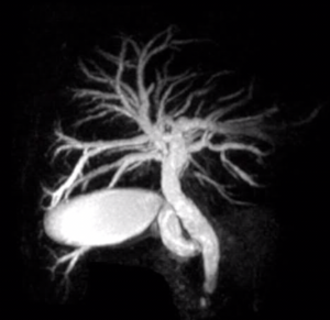
MRCP
Cardiac MR
The intricate anatomy and physiologic motion make cardiac evaluation the final frontier and the true test of any imaging equipment. Our MRI system and its software are capable of passing this test with flying colours. Internationally, Cardiac MR is now considered the gold standard for detection of myocardial infarction, scarring, cardiomyopathies and to assess myocardial viability. It is also useful in valvular assessment in special cases as an adjunct to echocardiography. Explore our services for Cardiac MRI scan in Mumbai with competitive pricing.


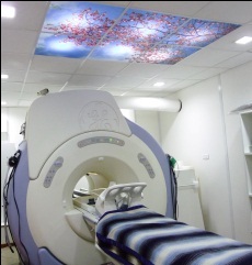
Daylighting system in MR room
Provides a bright ambience which minimises claustrophobia.
