Introduction
A PET scan, short for Positron Emission Tomography, is a powerful imaging test that helps doctors see how your organs and tissues are functioning. Unlike X-rays or CT scans, which only show the structure, a PET scan can detect changes at the cellular level, making it a vital tool for early diagnosis.
Whether you are concerned about cancer, heart conditions, or neurological disorders, understanding what a PET scan can do helps you make informed healthcare decisions. This guide explains everything you need to know, from what a PET scan detects to how it works and when it’s recommended.

What is a PET Scan?
A PET scan is a non invasive imaging test that reveals how your body’s organs and tissues are working. It uses a small amount of radioactive material, called a tracer, to highlight areas of increased chemical activity in the body.
How it differs from other scans
- CT/MRI scans: Show detailed images of structures but not function.
- PET scan: Shows metabolic activity, which helps detect disease earlier than structural changes appear.
PET scans are commonly used in oncology, cardiology, and neurology to provide precise insights that other scans may miss.
How Does a PET Scan Work?
Here’s a step-by-step overview of the PET scan process:
- Preparation
- You may need to fast for 4–6 hours before the scan.
- Certain medications may need to be paused.
- Avoid strenuous activity 24 hours prior.
- You may need to fast for 4–6 hours before the scan.
- Injection of Tracer
- A small dose of radioactive tracer is injected into a vein.
- The tracer travels through your bloodstream and accumulates in active tissues.
- A small dose of radioactive tracer is injected into a vein.
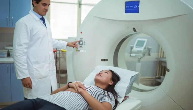
- Scanning Process
- You lie on a cushioned table that slides into a PET scanner.
- The scanner detects the tracer and creates detailed images of your body’s metabolic activity.
- The scan usually takes 30–60 minutes.
- You lie on a cushioned table that slides into a PET scanner.
- Post-Scan
- You can resume normal activities immediately.
- Drink plenty of water to help flush the tracer from your body.
- You can resume normal activities immediately.
PET scans are painless and generally safe, with minimal radiation exposure.
What Can a PET Scan Detect?
PET scans are versatile and can detect a variety of conditions:
- Cancer Detection and Monitoring
- Identifies tumors and metastases.
- Monitors treatment effectiveness.
- Heart Conditions
- Detects areas of reduced blood flow.
- Assesses damage after a heart attack.
- Brain Disorders
- Detects Alzheimer’s disease, epilepsy, and dementia.
- Helps plan neurological treatments.
- Other Uses
- Detects infections and inflammation.
When Do You Need a PET Scan?
A PET scan may be recommended if you have:
Doctors often use PET scans alongside other tests like CT or MRI to get a complete picture of your health.
- Persistent unexplained pain or symptoms.
- A history of cancer for diagnosis, staging, or monitoring.
- Heart problems require assessment of blood flow or damage.
- Neurological symptoms like seizures or memory loss.
- Unexplained infections or inflammatory conditions.
Benefits of a PET Scan
- Early detection: Identifies diseases before structural changes occur.
- Non-invasive: No surgery or complicated procedures needed.
- Accurate: Provides detailed metabolic information.
Treatment planning: Helps doctors choose the best therapy.
Preparing for a PET Scan
- Fasting: Usually 4–6 hours before the scan.
- Medications: Inform your doctor about any drugs you are taking.
- Hydration: Drink water before and after the scan.
Frequently Asked Questions (FAQs)
1. What is the difference between a PET scan and a CT scan?
CT shows structure; PET shows function and metabolic activity.
2. Is a PET scan safe?
Yes, it involves a small amount of radiation, which is generally safe.
3. How long does a PET scan take?
Typically 30 to 60 minutes, plus preparation time.
4. Can PET scans detect cancer early?
Yes, PET scans can identify cellular changes before tumors become visible on other scans.
5. Will I feel pain during the scan?
No, the scan is painless; you only feel a small injection.
Conclusion
A PET scan is a valuable diagnostic tool that goes beyond structural imaging to reveal how your body is functioning at a cellular level. From detecting cancer and heart disease to neurological conditions, it provides critical insights that guide treatment decisions.
If you or a loved one may benefit from a PET scan, consult your doctor to discuss whether it’s the right choice. Early detection can make a life-saving difference.
At Ace Imaging Centre, we combine advanced technology, expert radiologists, and a patient first approach to deliver accurate and safe imaging results that help your doctor plan the best treatment for you.
Name: Ace Imaging Center
Address: Bezolla Complex, 102, VN Purav Marg, Swastik Park, Chembur, Mumbai, Maharashtra 400071
contact: 081693 60334