DOTA scan in Mumbai

DOTA Scan in Mumbai: Benefits, Process, and When It’s Needed

In the realm of advanced medical imaging, DOTA scan in Mumbai has emerged as a pivotal tool for diagnosing and managing various health conditions. Conducted at leading facilities such as Ace Imaging, this procedure provides critical insights into the body’s functional processes. In this blog, we will delve into the benefits, the process, and the specific circumstances under which a DOTA scan is recommended.

What is a DOTA Scan?

A DOTA (Diethylene Triamine Pentaacetic Acid) scan, often referred to as a somatostatin receptor scintigraphy, is a specialized imaging technique that utilizes a radioactive tracer to visualize specific cellular functions in the body. This scan is particularly effective in identifying neuroendocrine tumors and other conditions related to hormone-secreting cells.

Benefits of DOTA Scan

1. Early Detection of Diseases

One of the primary advantages of a DOTA scan is its ability to detect diseases at an early stage. This is crucial for conditions like neuroendocrine tumors, where timely intervention can significantly improve patient outcomes.

2. Non-Invasive Procedure

The DOTA scan is a non-invasive imaging technique, meaning it does not require any surgical procedures. Patients can undergo the scan comfortably without the need for anesthesia or extensive preparation.

3. Comprehensive Imaging

DOTA scans provide comprehensive imaging that helps in visualizing not just tumors but also the functionality of organs and tissues. This comprehensive view is essential for accurate diagnosis and treatment planning.

4. Minimal Side Effects

The procedure has minimal side effects compared to other diagnostic tests. Most patients can return to their normal activities shortly after the scan, making it a convenient option for many.

The Process of a DOTA Scan

Preparation

Before undergoing a DOTA scan, patients may be advised to follow certain preparatory steps, which could include:

  • Fasting: Patients are often required to fast for a few hours before the scan.
  • Medications: Inform your doctor about any medications you are currently taking, as some may need to be paused before the scan.

Procedure

The DOTA scan involves the following steps:

  1. Injection of Tracer: A radioactive tracer is injected into a vein, typically in the arm. This tracer binds to the somatostatin receptors present in certain tumors and tissues.
  2. Waiting Period: After the injection, there is usually a waiting period of about 1 to 4 hours to allow the tracer to circulate and accumulate in the target areas.
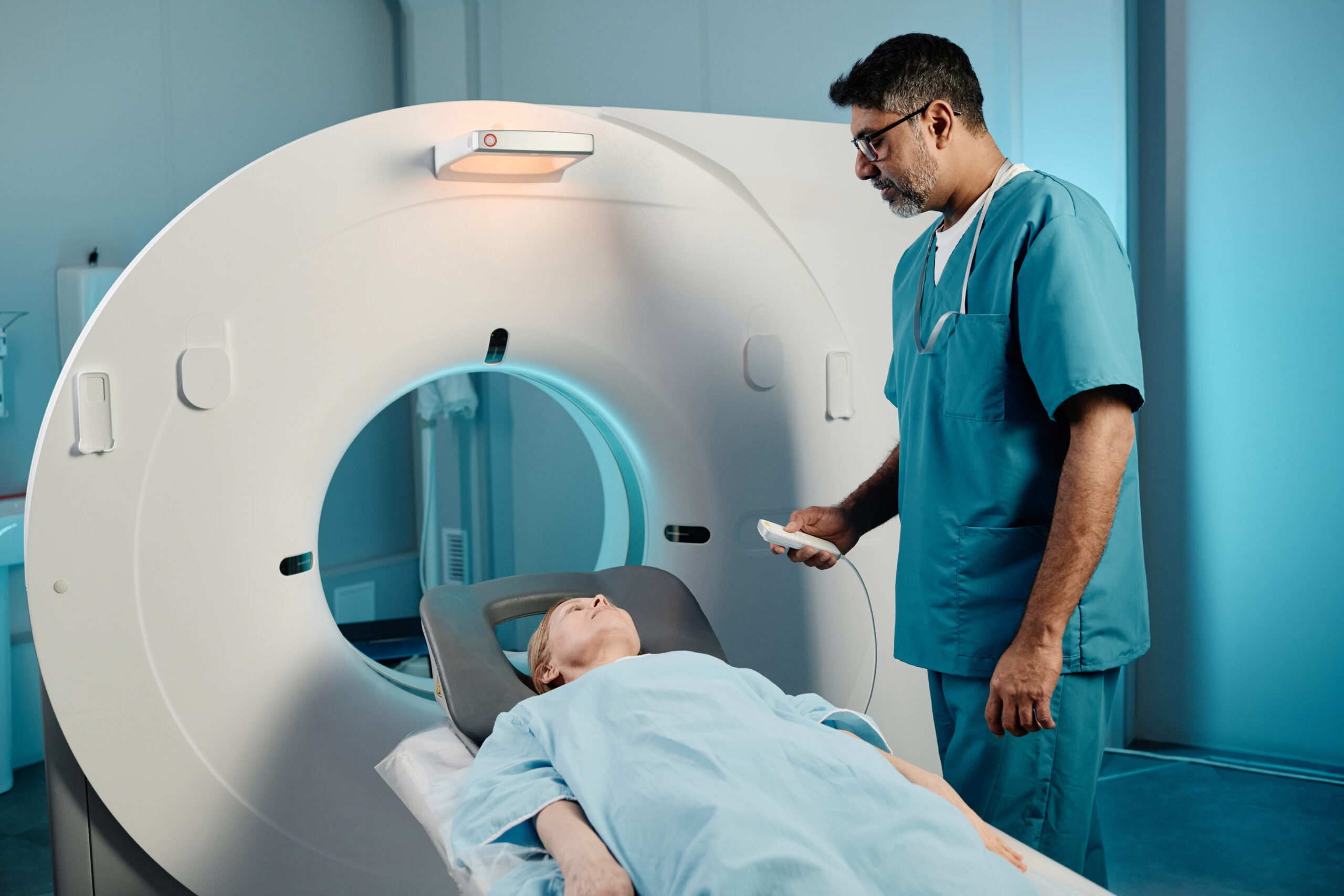
  3. Imaging: The patient will then be positioned in front of a gamma camera that detects the radiation emitted by the tracer. Multiple images will be taken from various angles to create detailed pictures of the body.
  4. Duration: The entire process, including the waiting time, can take several hours, but the actual imaging usually lasts about 30 minutes.

When is a DOTA Scan Needed?

A DOTA scan is typically recommended in the following situations:

  • Suspicion of Neuroendocrine Tumors: If a doctor suspects a neuroendocrine tumor based on symptoms or initial tests, a DOTA scan can confirm the diagnosis.
  • Monitoring Treatment Response: Patients undergoing treatment for neuroendocrine tumors may need periodic DOTA scans to assess how well the treatment is working.
  • Evaluating Symptoms: Unexplained hormonal symptoms may lead physicians to recommend a DOTA scan to check for potential underlying causes.

Conclusion

If you’re seeking a DOTA scan in Mumbai, Ace Imaging offers state-of-the-art facilities and expert medical staff to ensure a smooth experience. With its ability to provide vital information about neuroendocrine tumors and other conditions, the DOTA scan is an invaluable tool in modern medical diagnostics.

For more information about the DOTA scan process, benefits, or to schedule an appointment, feel free to contact Ace Imaging today!

MRI Scan in Mumbai

MRI Scan in Mumbai: Understanding the Process, Benefits, and Cost

What is an MRI Scan?

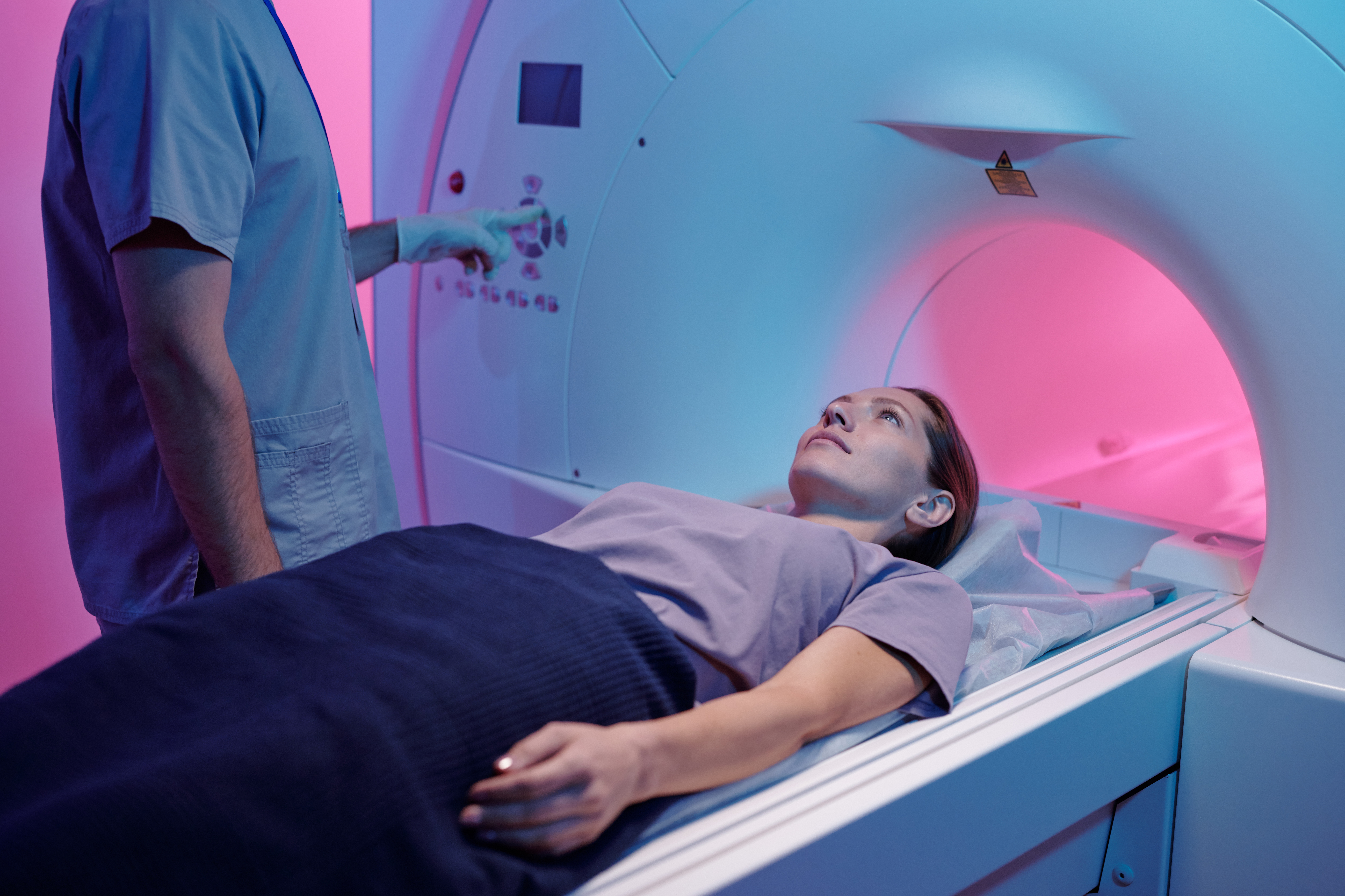
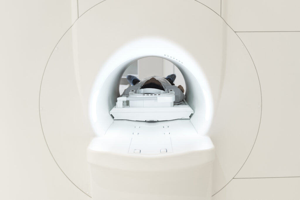
Magnetic Resonance Imaging (MRI) is a non-invasive imaging technique that uses powerful magnets and radio waves to create detailed images of the body’s internal structures. Unlike X-rays or CT scans, MRI scans do not involve radiation, making them safer for frequent use.

Why Choose an MRI Scan in Mumbai?

Mumbai is home to world-class medical facilities, including Ace Imaging, which is renowned for its cutting-edge technology and experienced radiologists. Opting for an MRI Scan in Mumbai ensures access to the latest diagnostic tools, accurate imaging, and reliable medical expertise.

Benefits of Getting an MRI Scan

  1. Detailed Imaging: MRI scans provide high-resolution images, which are ideal for detecting abnormalities in soft tissues, organs, and the nervous system.
  2. Non-Invasive and Safe: The procedure is non-invasive and does not use harmful radiation.
  3. Wide Range of Applications: MRI scans are versatile and used to diagnose various conditions, including brain injuries, spinal disorders, joint issues, and abdominal problems.
  4. Customized Scans: MRI technology can be adapted to focus on specific body areas, allowing for targeted diagnosis.

How is an MRI Scan Performed?

Getting an MRI Scan in Mumbai typically follows these steps:

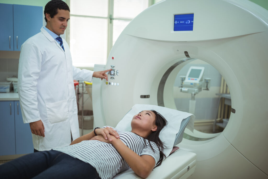
  1. Preparation: Patients may be asked to change into a hospital gown and remove any metal objects to avoid interference.
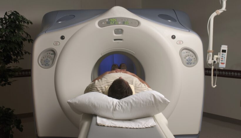
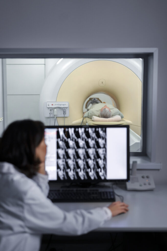
  2. Positioning: The patient lies on a motorized table that slides into the MRI machine.
  3. The Scanning Process: The machine produces loud thumping or tapping noises, and patients may wear ear protection. The scan can take anywhere from 15 minutes to over an hour, depending on the body part being examined.
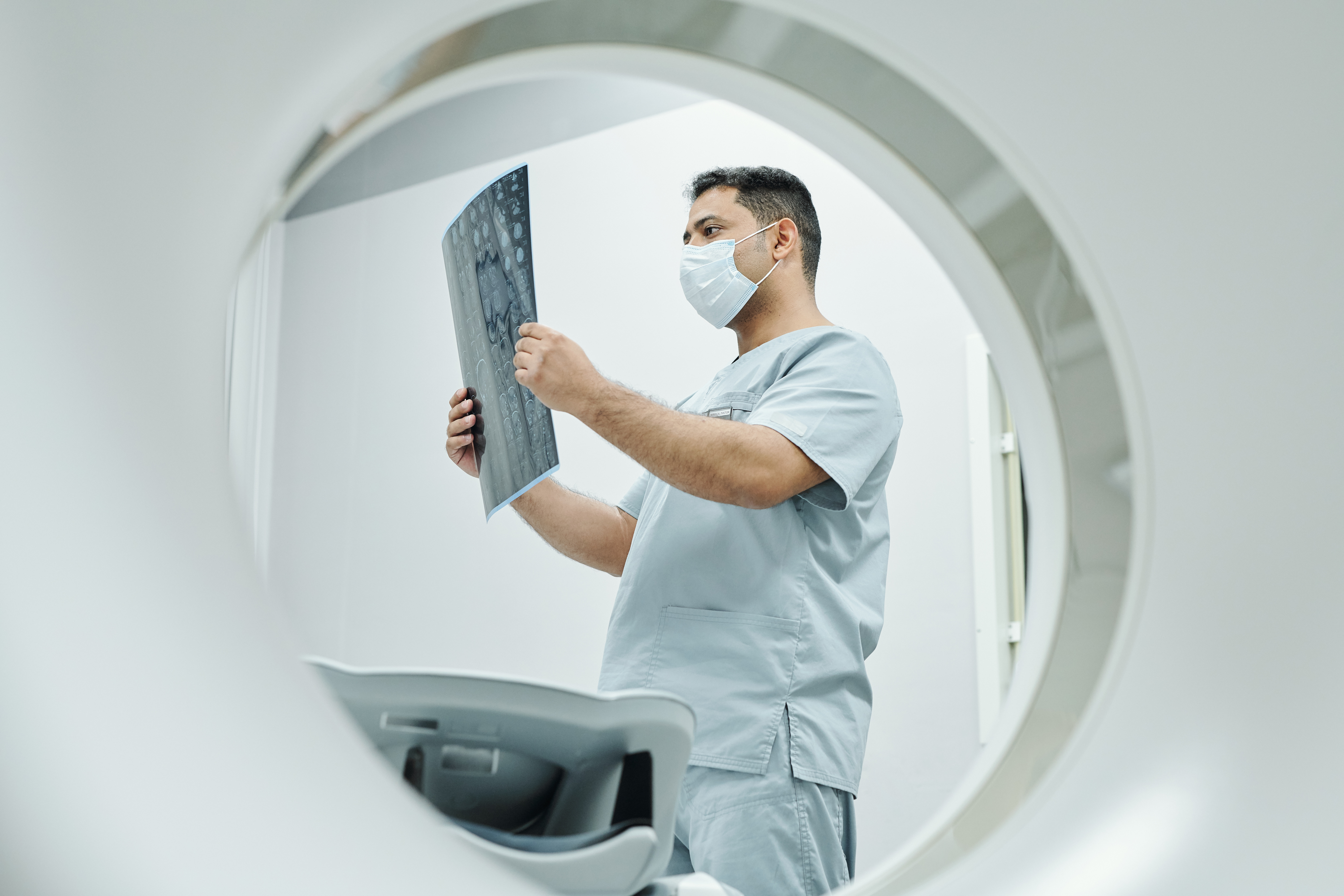
  4. Imaging Results: Once the scan is complete, radiologists at Ace Imaging review the images and create a detailed report for your doctor.

Cost of an MRI Scan in Mumbai

The cost of an MRI Scan in Mumbai varies depending on several factors, including the type of scan, the facility, and any additional procedures involved. At Ace Imaging, patients benefit from transparent pricing and high-quality service. Generally, MRI scan prices range from INR 5,000 to INR 15,000 or more, depending on complexity.

Factors Affecting MRI Scan Costs

  • Type of MRI: Advanced scans, such as contrast-enhanced MRIs, are priced higher.
  • Body Part: Scans for specific areas like the brain or spine might differ in cost.
  • Technology and Equipment: Facilities with state-of-the-art machines may charge more for their services.
  • Expertise of the Radiologist: Experienced radiologists who provide detailed and accurate interpretations may contribute to higher costs.

Why Choose Ace Imaging for Your MRI Scan?

Ace Imaging stands out for several reasons:

  • Advanced MRI Technology: Cutting-edge equipment ensures high-quality images.
  • Experienced Professionals: Skilled radiologists and technicians with extensive experience in diagnostic imaging.
  • Patient-Centric Care: The facility prioritizes comfort and efficient service.

FAQs About MRI Scans in Mumbai

Q1: Is an MRI scan painful?
No, MRI scans are painless. However, some patients may experience discomfort from staying still for an extended period.

Q2: Are there any risks associated with an MRI?
MRI scans are generally safe. The primary concern is for patients with certain implants, such as pacemakers, as the magnetic field may interfere with them.

Q3: How should I prepare for my MRI scan at Ace Imaging?
You may be instructed to avoid food and drink before your scan, especially if a contrast dye is used. Always inform your doctor and the radiologist of any metal implants or medical conditions.

Conclusion

Choosing an MRI Scan in Mumbai at Ace Imaging ensures a seamless diagnostic experience with expert care and cutting-edge technology. Understanding the process, benefits, and costs can help you make an informed decision about your health.

PET Scans for Early Cancer Detection in Mumbai

The Benefits of PSMA PET Scans for Early Cancer Detection in Mumbai

Introduction In the fight against cancer, early and accurate diagnosis is crucial. The PSMA PET Scan in Mumbai has become a groundbreaking tool for detecting prostate cancer and other cancers at an early stage. This article delves into the benefits of this advanced imaging technique and how Ace Imaging in Mumbai is leading the way in offering this vital service.

What is a PSMA PET Scan? PSMA PET Scan (Prostate-Specific Membrane Antigen Positron Emission Tomography) is an advanced imaging test that provides detailed pictures of the prostate and other tissues where cancer may spread. This technology helps doctors identify cancer cells earlier and more accurately than traditional scans.

How PSMA PET Scan Works The PSMA Scan works by injecting a small amount of a radioactive tracer that binds to PSMA proteins commonly found on prostate cancer cells. This tracer makes the cancer cells glow on the PET scan, providing doctors with precise information about the location and extent of cancer.

Advantages of PSMA Scan for Early Cancer Detection 1. Enhanced Detection Capabilities The PSMA PET Scan in Mumbai stands out for its ability to detect even small cancerous lesions that other imaging techniques might miss. This is crucial for initiating early treatment.

  1. Accurate Staging and Treatment Planning For patients diagnosed with prostate cancer, knowing the exact stage is essential for effective treatment. Ace Imaging provides PSMA PET Scans that help map out the cancer’s reach, ensuring that doctors can create targeted treatment plans.
  2. Reduced Need for Multiple Tests Traditional imaging might require multiple tests to confirm cancer presence and spread. A PSMA Scan consolidates this process, offering comprehensive insights in a single session, saving both time and cost.

Why Choose PSMA Scan Over Other Imaging Techniques?

  • Higher Sensitivity and Specificity: The scan’s ability to identify cancerous tissues with greater sensitivity than CT or MRI scans.
  • Less Invasive: While being highly informative, a PSMA Scan is non-invasive and well-tolerated by patients.
  • Efficient Diagnosis: It provides quicker and more reliable results, which helps reduce the anxiety of waiting for multiple tests.

The Role of Ace Imaging in Delivering Quality PSMA PET Scans in Mumbai Ace Imaging in Mumbai is at the forefront of providing state-of-the-art diagnostic imaging services. Equipped with the latest technology and a team of skilled radiologists, Ace Imaging ensures that patients receive accurate, timely, and compassionate care.

What Sets Ace Imaging Apart?

  • Cutting-edge Technology: Utilizing the latest advancements in PET scan technology.
  • Experienced Team: Radiologists with specialized training in interpreting PSMA PET Scans.
  • Comprehensive Care: A patient-centered approach that emphasizes clear communication and tailored care plans.

Conclusion The PSMA PET Scan in Mumbai offered by Ace Imaging is revolutionizing early cancer detection. With unmatched precision, faster results, and enhanced detection capabilities, this advanced imaging technique is invaluable for patients and healthcare providers alike. Choosing Ace Imaging for a PSMA Scan ensures you receive the highest standard of diagnostic care, contributing to better outcomes and peace of mind.

Mammography

Mammography – advances and safety of screening procedures

Mammography, a key tool in breast cancer screening, has undergone significant advancements in technology and safety measures over the years. 

Advances in Mammography Technology:

Digital Mammography: Digital mammography has largely replaced conventional film-screen mammography, offering improved image quality, faster image acquisition, and digital storage capabilities. This technology allows for better visualization of breast tissue, particularly in women with dense breasts.

3D Mammography (Tomosynthesis): Three-dimensional (3D) mammography, also known as tomosynthesis, provides multiple thin-slice images of the breast, reducing overlapping tissue and improving cancer detection rates. 

Contrast-Enhanced Mammography: Contrast-enhanced mammography involves the use of intravenous contrast agents to improve the visualization of blood flow within breast lesions. This technique enhances the detection of suspicious areas and aids in characterizing breast abnormalities.

Automated Breast Ultrasound (ABUS): ABUS is a supplementary imaging modality used in women with dense breasts to improve the detection of breast cancers that may be obscured on mammography. It provides a detailed evaluation of breast tissue without exposing patients to ionizing radiation.

Safety Considerations in Mammography Screening:

Radiation Exposure: Mammography uses ionizing radiation to produce images of the breast. While the radiation dose from mammography is low and considered safe, efforts are made to minimize radiation exposure without compromising image quality. Modern digital mammography systems are designed to optimize radiation dose levels.

Compression Discomfort: Mammography requires breast compression to spread out breast tissue and obtain clear images. While breast compression can cause discomfort for some women, it is necessary to improve image quality and reduce motion artifacts. Technologists strive to minimize discomfort while ensuring adequate compression.

Breast Density Awareness: Breast density, a measure of the proportion of glandular and connective tissue relative to fat in the breast, can affect the accuracy of mammography. Women with dense breasts may benefit from additional screening modalities such as tomosynthesis or ultrasound to improve cancer detection rates.

Overall, mammography remains an essential tool in breast cancer screening, with ongoing advancements in technology and safety measures aimed at improving early detection and reducing the burden of breast cancer. Regular mammography screening, combined with clinical breast exams and self-breast awareness, plays a critical role in early detection and improving outcomes for women at risk of breast cancer.