Radiology field in the next decade – a game changer

The radiology field is poised for several transformative developments in the coming decade, with advancements in technology, artificial intelligence (AI), and personalized medicine driving significant changes. Here are some potential game changers:

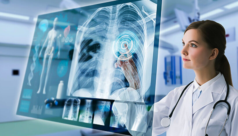
AI and Machine Learning Integration: AI algorithms will continue to revolutionize radiology by enhancing image interpretation, improving accuracy, and expediting diagnosis. These technologies will assist radiologists in detecting abnormalities, prioritizing cases, and providing quantitative analysis, ultimately leading to more efficient and precise patient care.

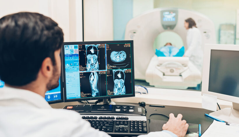
Quantitative Imaging: There will be a shift towards quantitative imaging techniques that provide more objective and reproducible measurements of disease characteristics, such as tumor volume, perfusion, and metabolic activity. This will enable better treatment planning, response assessment, and prognostication in conditions like cancer, cardiovascular disease, and neurological disorders.

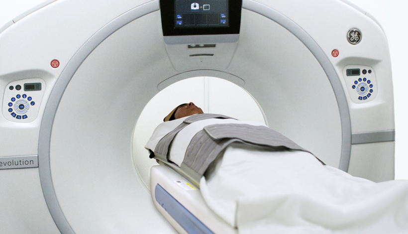
Advancements in Imaging Modalities: Emerging imaging modalities, such as molecular imaging, spectral CT, and functional MRI, will offer deeper insights into biological processes at the cellular and molecular levels. These technologies will facilitate early detection, characterization, and treatment monitoring of various diseases, leading to improved patient outcomes.

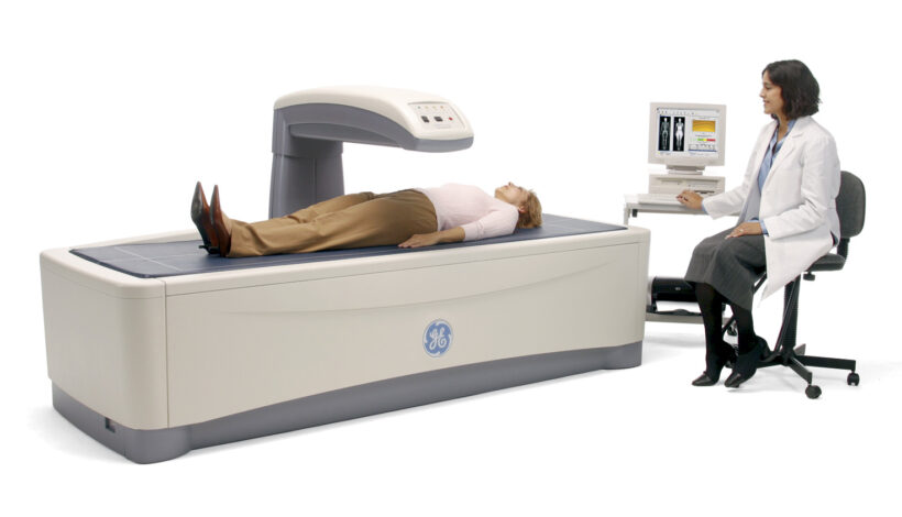
Point-of-Care Imaging: Portable and handheld imaging devices will become more prevalent, allowing for rapid and convenient imaging at the bedside or in remote locations. These point-of-care devices will facilitate timely diagnosis and decision-making, particularly in emergency and critical care settings.

Precision Radiology and Personalized Medicine: Radiomics, radiogenomics, and other precision medicine approaches will enable the extraction of valuable quantitative data from medical images, leading to personalized treatment strategies tailored to individual patient characteristics and disease biology.

Enhanced Collaboration and Integration: Radiology will increasingly collaborate with other medical specialties, such as pathology, genomics, and molecular biology, to provide comprehensive diagnostic and therapeutic solutions. Interdisciplinary teams will work together to integrate imaging findings with clinical data, leading to more holistic patient management.

Patient-Centric Care: There will be a greater emphasis on patient-centered imaging, focusing on safety, comfort, and communication. Radiologists will play a more active role in patient education, shared decision-making, and follow-up care, ensuring a positive and empowering experience for patients undergoing imaging examinations.

Overall, the next decade promises to be an exciting and transformative period for the radiology field, with innovations in technology, AI, and personalized medicine driving improvements in diagnosis, treatment, and patient care.

Radiology

Radiology field : booming or obsolete

The radiology field is far from obsolete; in fact, it continues to evolve and thrive, driven by technological advancements, demographic changes, and increasing healthcare needs. Here’s why the radiology field is booming:

Technological Advancements: Radiology is at the forefront of technological innovation, with advancements in imaging modalities, image processing software, and artificial intelligence transforming the field. Technologies such as MRI, CT, PET-CT, and digital mammography are continuously improving, enabling more accurate diagnosis, treatment planning, and monitoring of diseases.

Growing Demand: The demand for radiology services is increasing due to several factors, including an aging population, rising incidence of chronic diseases, and advances in preventive medicine. Radiology plays a crucial role in the early detection, diagnosis, and management of various conditions, from cancer and cardiovascular disease to neurological disorders and musculoskeletal injuries.

Expanded Applications: Radiology is expanding beyond traditional diagnostic imaging to include interventional radiology, nuclear medicine, molecular imaging, and image-guided therapies. These subspecialties offer minimally invasive treatment options and personalized approaches to patient care, further driving demand for radiology services.

Role of Artificial Intelligence: Artificial intelligence (AI) is revolutionizing radiology by enhancing image interpretation, workflow efficiency, and diagnostic accuracy. AI algorithms can assist radiologists in detecting abnormalities, prioritizing cases, and predicting patient outcomes, ultimately improving patient care and outcomes.

Global Health Challenges: Radiology plays a critical role in addressing global health challenges such as infectious diseases, pandemics, and public health emergencies. Imaging techniques are essential for early detection and surveillance of infectious agents, monitoring disease progression, and guiding treatment strategies.

Interdisciplinary Collaboration: Radiology is increasingly integrated into multidisciplinary care teams, collaborating with clinicians, surgeons, oncologists, and other healthcare professionals to provide comprehensive and coordinated patient care. This collaborative approach enhances patient outcomes and fosters innovation in research and clinical practice.

Career Opportunities: The radiology field offers diverse career opportunities for healthcare professionals, including radiologists, radiologic technologists, radiation therapists, medical physicists, and radiology nurses. With ongoing advancements and specialization within the field, there are abundant opportunities for professional growth and development.

Overall, the radiology field is thriving and poised for continued growth, driven by technological innovation, expanding applications, and the increasing importance of imaging in modern healthcare. Far from being obsolete, radiology remains indispensable in the diagnosis, treatment, and management of a wide range of medical conditions, contributing to improved patient outcomes and quality of care.

LDCT scan in mumbai

Low dose CT : new technology for lung cancer screening – is it really worth?CTLow dose CT : new technology for lung cancer screening – is it really worth?

Low-dose computed tomography (LDCT) has gained attention as a screening tool for lung cancer, particularly in individuals at high risk for the disease. The use of LDCT for lung cancer screening has been studied extensively, and it has shown promise in detecting lung cancers at earlier, more treatable stages. 

Advantages of LDCT for Lung Cancer Screening:

Early Detection: LDCT has demonstrated a higher sensitivity in detecting small lung nodules compared to traditional chest X-rays. Early detection can lead to more effective treatment and improved outcomes.

Reduced Mortality: Clinical trials, such as the National Lung Screening Trial (NLST), have shown that lung cancer screening with LDCT can lead to a reduction in lung cancer mortality compared to chest X-ray screening.

Risk Stratification: LDCT is generally recommended for individuals at high risk for lung cancer, such as those with a significant smoking history. This helps in targeting the screening to those who are more likely to benefit.

Less Invasive than Biopsy: Detecting lung nodules early through LDCT may reduce the need for more invasive diagnostic procedures like lung biopsies.

Improved Survival Rates: Early-stage lung cancers often have better prognosis and treatment options. LDCT screening may contribute to improved survival rates by identifying cancers before they advance.

Pitfalls: 

False Positives:  LDCT may lead to false-positive results, where nodules are detected that are not cancerous. This can lead to additional tests, potential anxiety, and unnecessary interventions.

Radiation Exposure: Although LDCT uses lower radiation doses compared to standard CT scans, it still involves exposure to ionizing radiation. The balance between benefits and potential harm from radiation should be considered.

Appropriate Patient Selection: Identifying individuals at high risk for lung cancer is crucial for the effectiveness of LDCT screening. Screening should be targeted toward those who are most likely to benefit.

Recommendations:

The U.S. Preventive Services Task Force (USPSTF) recommends annual LDCT screening for individuals aged 50-80 years who have a 20 pack-year smoking history and currently smoke or have quit within the past 15 years.

Shared decision-making between patients and healthcare providers is essential to discuss the potential benefits and risks of LDCT screening based on individual factors.

In conclusion, LDCT for lung cancer screening can be valuable, especially for high-risk individuals, as it has the potential to detect lung cancer at earlier stages and reduce mortality. However, the decision to undergo screening should be individualized, considering the specific risks and benefits for each person. 

DEXA

Decoding DEXA scans : Understanding bone density assessment

Dual-energy X-ray absorptiometry (DEXA) scans are a commonly used diagnostic tool to assess bone density and are particularly valuable in diagnosing and monitoring conditions such as osteoporosis. Let’s break down the key aspects of decoding DEXA scans and understanding bone density assessment:

Principle of DEXA: DEXA uses two X-ray beams, each with different energy levels, to measure the absorption of these beams by bone and soft tissue. The attenuation of the X-ray beams by bone is directly related to bone mineral density (BMD).

Bone Mineral Density (BMD):

BMD is a measure of the amount of mineral (mostly calcium and phosphorus) per unit volume in bone. DEXA measures BMD at various skeletal sites, commonly the spine, hip, and forearm.

T-score and Z-score:

T-score: This score compares an individual’s BMD with that of a healthy young adult of the same sex. It is used in diagnosing osteoporosis.

T-score between +1 and -1: Normal

T-score between -1 and -2.5: Osteopenia (low bone mass)

T-score -2.5 or lower: Osteoporosis

Z-score: This score compares BMD with the average BMD of individuals of the same age, sex, and ethnicity.

Z-score below -2: Lower than expected for age (may indicate secondary causes of bone loss)

Regions of Interest (ROI): DEXA scans typically assess specific regions, including the lumbar spine, femoral neck, and total hip. These regions are prone to fractures and are crucial for diagnosing osteoporosis.

Osteoporosis and Fracture Risk: Osteoporosis is characterized by low bone mass and structural deterioration, leading to an increased risk of fractures. DEXA scans help identify individuals at risk for fractures, allowing for early intervention to prevent bone loss and fractures.

Follow-up and Monitoring: DEXA scans are used for monitoring changes in bone density over time, evaluating the effectiveness of treatment, and adjusting therapeutic approaches accordingly.

Factors Influencing BMD: Various factors, including age, sex, genetics, hormonal status, and lifestyle factors (nutrition, physical activity, smoking), influence BMD.

Clinical Recommendations: DEXA screening is recommended for postmenopausal women and older men with risk factors for fractures. The frequency of DEXA scans depends on individual risk factors and initial findings. Understanding DEXA scan results requires collaboration between patients and healthcare providers to interpret the findings in the context of overall health and risk factors. Regular assessments help in early detection and effective management of bone-related conditions.