PSMA pet scan in Mumbai

PSMA PET Scan: A Game Changer for Prostate Cancer Patients in Mumbai

Prostate cancer is one of the most common types of cancer affecting men, with millions of new cases diagnosed worldwide each year. In India, and particularly in metropolitan cities like Mumbai, the prevalence of prostate cancer is steadily rising. Early detection and accurate staging of prostate cancer are critical for determining the most effective treatment approach. In this context, the PSMA PET scan in Mumbai has emerged as a revolutionary tool in the diagnosis and management of prostate cancer. For patients in Mumbai, understanding the benefits of this cutting-edge technology can make a significant difference in their treatment outcomes.

What is a PSMA PET Scan?

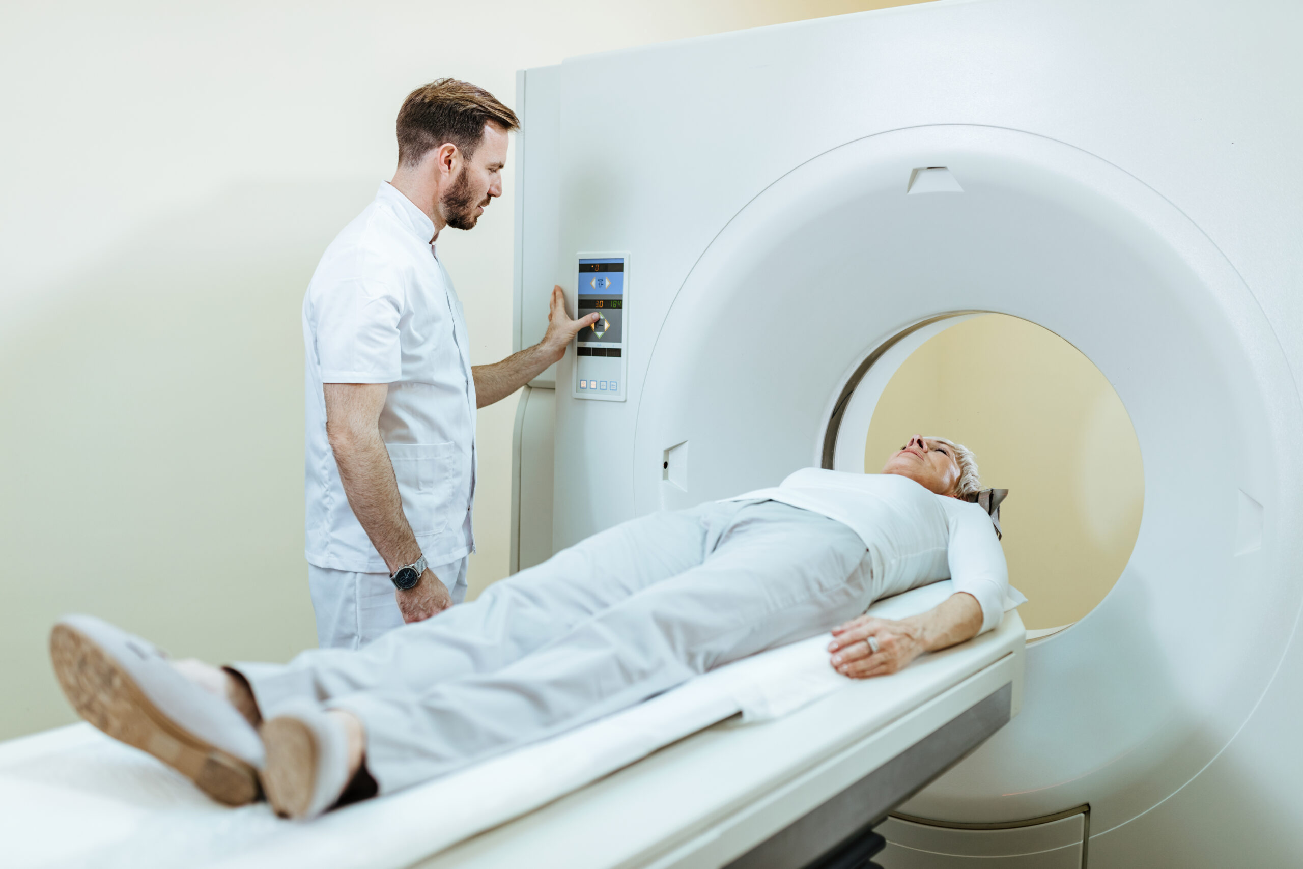
The PSMA PET scan (Prostate-Specific Membrane Antigen Positron Emission Tomography) is a highly advanced imaging technique used to detect prostate cancer. It uses a specialized radioactive tracer that binds to the PSMA protein, which is present in higher concentrations on the surface of prostate cancer cells. Once injected into the patient, the tracer allows doctors to visualize prostate cancer cells more clearly during the PET scan, helping to identify tumors that may not be visible with conventional imaging techniques like CT or MRI.

This scan provides a more detailed and accurate picture of the prostate cancer’s spread, making it easier for doctors to determine the extent of the disease. In addition to detecting primary tumors, the PSMA PET scan can also identify small metastases or recurrent cancer cells in distant organs, which may otherwise go undetected.

Why is the PSMA PET Scan a Game Changer for Prostate Cancer?

  1. Improved Detection of Prostate Cancer

Traditionally, the diagnosis and staging of prostate cancer have relied on imaging techniques like CT scans, MRI, and bone scans. While these methods are useful, they often miss small, early-stage tumors or metastases. The PSMA PET scan in Mumbai, however, is much more sensitive and specific when it comes to detecting prostate cancer cells. This means it can help detect the disease at an earlier stage, giving patients a better chance of receiving timely and targeted treatment.

For men in Mumbai, where the risk of prostate cancer is on the rise, the ability to detect prostate cancer early can dramatically improve survival rates and quality of life. Early detection can lead to more treatment options and a less invasive approach, making the PSMA PET scan an invaluable tool for proactive care.

  1. Accurate Staging and Localization

One of the most significant advantages of a PSMA PET scan over traditional imaging techniques is its ability to provide detailed and precise staging information. For prostate cancer patients, knowing the exact location and extent of cancer spread is crucial for deciding the most effective treatment plan. Whether the cancer has spread to nearby lymph nodes, bones, or distant organs, the PSMA PET scan can pinpoint the location of metastases, helping doctors tailor a treatment plan based on the specific areas involved.

This level of accuracy allows for a more personalized treatment approach. For example, if the scan shows that the cancer has spread to a particular area, doctors can focus on that region with targeted radiation therapy or other treatments. The PSMA PET scan ensures that the treatment plan is as specific and effective as possible, increasing the chances of success.

  1. Better Detection of Recurrence

Prostate cancer can often recur after initial treatment, especially if small cancer cells were missed during the first round of scans. The PSMA PET scan is particularly effective in detecting recurrent prostate cancer, even in cases where traditional imaging methods have failed. This is crucial for patients who have already undergone surgery, radiation therapy, or hormone therapy and are being monitored for signs of cancer returning.

For Mumbai patients, this means that the PSMA PET scan can help identify recurrence early, allowing for prompt intervention and more effective treatment. Detecting recurrence at an early stage improves the likelihood of successful second-line treatments, which can significantly extend life expectancy and quality of life for prostate cancer patients.

  1. Non-Invasive and Convenient

Unlike biopsy procedures, which can be invasive and require recovery time, the PSMA PET scan is a non-invasive diagnostic tool. Patients in Mumbai benefit from the fact that it offers a detailed and accurate diagnosis without the need for surgical intervention. The scan itself is quick and typically requires no special preparation, making it a convenient option for patients who need to monitor their prostate cancer status.

Additionally, since the PSMA PET scan is performed in a single session, it eliminates the need for multiple diagnostic tests, providing a quicker and more efficient way to evaluate the extent of the cancer.

  1. A More Personalized Approach to Treatment

The primary advantage of the PSMA PET scan lies in its ability to provide a more personalized treatment approach for prostate cancer patients. Based on the scan results, doctors can select the most appropriate treatment options, which may include surgery, radiation therapy, hormone therapy, or chemotherapy. For patients with advanced or metastatic prostate cancer, the PSMA PET scan may help identify specific areas for targeted therapies, potentially improving treatment outcomes and minimizing side effects.

For men in Mumbai who are diagnosed with prostate cancer, having access to this advanced imaging technology means they are more likely to receive care tailored to their specific needs. Whether they are in the early stages of the disease or have advanced metastatic cancer, the PSMA PET scan allows for treatment decisions that are based on precise, up-to-date information about the cancer’s location and spread.

Why Should Mumbai Patients Consider a PSMA PET Scan?

  1. Access to Advanced Healthcare Facilities

Mumbai is home to some of India’s leading hospitals and diagnostic centers, which offer the latest in medical technology. With the availability of PSMA PET scans at top medical centers, patients in Mumbai have access to world-class prostate cancer care. The growing availability of this diagnostic tool means that more men in the city can benefit from accurate, early detection and personalized treatment plans.

  1. Growing Prostate Cancer Awareness

As awareness of prostate cancer continues to grow in Mumbai and across India, more men are becoming proactive about their health and seeking diagnostic tests such as the PSMA PET scan. By considering this advanced imaging technique, patients can take charge of their prostate health and ensure that they receive the best possible care available.

  1. A Better Chance of Beating the Disease

Ultimately, the earlier prostate cancer is detected and treated, the better the chances of successful outcomes. The PSMA PET scan in Mumbai is a game changer in this regard, providing detailed imaging that can catch even the most subtle signs of the disease. By offering a clearer picture of prostate cancer’s location and extent, it empowers doctors to make more informed decisions, improving treatment options and overall survival rates.

PSMA pet scan in Mumbai

Frequently Asked Questions (FAQs)

Q1: What is a PSMA PET scan?

A: A PSMA PET scan detects prostate cancer using a radioactive tracer that binds to cancer cells.

Q2: How is it different?

A: It’s more accurate than CT or MRI in detecting early cancer and metastases.

Q3: Is it safe?

A: Yes, it’s safe with minimal radiation.

Q4: How long does it take?

A: It takes 30 to 60 minutes.

Q5: Where can I get it in Mumbai?

A: Top hospitals and diagnostic centers in Mumbai offer PSMA PET scans.

Conclusion

For prostate cancer patients in Mumbai, the PSMA PET scan is a breakthrough in diagnosis and treatment. This cutting-edge imaging technology offers more accurate detection, better staging, and the ability to detect recurrence, all of which are crucial for effective treatment. As more hospitals and diagnostic centers in Mumbai adopt this innovative technique, patients now have access to the best tools for managing their prostate cancer.

If you or a loved one has been diagnosed with prostate cancer or is at risk, consider discussing the option of a PSMA PET scan with your healthcare provider. By utilizing this game-changing technology, patients can ensure they are taking the most effective steps in managing and treating prostate cancer, ultimately improving their chances of a positive outcome.

Pet Scan Centres in Mumbai

The Role of PET Scans in Modern Healthcare: A Comprehensive Guide

Positron Emission Tomography (PET) scans are an incredibly powerful diagnostic tool in modern healthcare. These scans help doctors and medical professionals gain detailed insights into the functioning of organs and tissues in the body, often detecting diseases or abnormalities much earlier than other imaging techniques. With increasing applications in diagnosing cancer, heart disease, neurological conditions, and more, PET scans are becoming an indispensable part of modern medicine.

What is a PET Scan?

A PET scan is a type of nuclear medicine imaging technique that provides detailed, three-dimensional images of the body. Unlike traditional imaging methods such as X-rays or CT scans, which primarily show the structure of the body, PET scans focus on the biochemical processes within tissues and organs.

The procedure involves injecting a small amount of a radioactive substance (a tracer) into the body. This tracer is typically a form of glucose or another molecule that is metabolized by cells. The PET scanner detects the radiation emitted by the tracer, allowing doctors to observe how the body is functioning at a cellular level. This makes it an essential tool for diagnosing and monitoring a wide range of medical conditions.

How Does a PET Scan Work?

  1. Tracer Injection: A radioactive tracer is injected into your body, usually through an IV. The tracer is a substance that emits positrons as it decays, and it’s designed to be absorbed by active tissues in the body.
  2. Scan Process: The PET scanner detects the radiation emitted by the tracer and creates detailed images based on this data. These images show how the tracer is distributed throughout your body, revealing active areas of metabolism and function.
  3. Data Analysis: The resulting images are then analyzed by radiologists and specialists to assess the presence of abnormalities, such as tumors, inflammation, or irregular blood flow.

Common Uses of PET Scans

  1. Cancer Diagnosis and Monitoring: One of the primary uses of PET scans is in cancer diagnosis. PET scans can detect the presence of cancerous cells by showing areas of high metabolic activity, which is often indicative of tumors. This is especially useful in identifying cancers that may not yet have visible structural changes (such as in the case of early-stage cancer). PET scans are also used to monitor how well a cancer treatment is working by assessing changes in the metabolic activity of a tumor.
  1. Cardiac Health: PET scans are also used to evaluate the heart’s function and blood flow. For patients with suspected coronary artery disease or other heart conditions, a PET scan can help doctors determine the health of the heart tissue, assess damage from previous heart attacks, and evaluate the effectiveness of treatments or procedures.
  2. Neurological Disorders: In the field of neurology, PET scans are valuable for diagnosing and monitoring conditions such as Alzheimer’s disease, epilepsy, and other neurological disorders. By assessing brain metabolism, PET scans can help detect changes in brain function that might indicate the presence of neurodegenerative diseases, even before symptoms become apparent.
  3. Infection and Inflammation: PET scans can also help detect infections or areas of inflammation in the body, which may be related to autoimmune disorders or other medical conditions. By identifying these areas, doctors can accurately determine the cause and location of the issue, enabling more targeted treatment.

Benefits of PET Scans

  • Early Detection: PET scans can detect changes in metabolic activity well before structural changes become evident in other imaging techniques. This allows for earlier detection of conditions like cancer or neurological diseases, leading to better outcomes.
  • Non-Invasive: While PET scans involve the use of radioactive tracers, the procedure itself is non-invasive. There are no incisions or needles beyond the initial injection of the tracer, making it relatively comfortable for patients.
  • Comprehensive Imaging: PET scans provide detailed, three-dimensional images of the body, allowing healthcare providers to observe both the structure and function of organs and tissues. This comprehensive information is invaluable for making accurate diagnoses and treatment plans.
  • Effective Monitoring: PET scans are useful for monitoring the progress of certain treatments, especially in cancer care. By showing how a tumor responds to treatment, doctors can make adjustments to the treatment plan as needed.

Potential Risks of PET Scans

While PET scans are generally safe, there are some risks associated with the procedure. These include:

  • Radiation Exposure: PET scans use radioactive tracers, which means there is some exposure to radiation. However, the levels used in PET scans are considered safe and are typically much lower than the risks posed by the conditions being diagnosed.
  • Allergic Reactions: Some patients may experience allergic reactions to the tracer, though this is rare.
  • Pregnancy and Breastfeeding: PET scans are typically avoided in pregnant women due to the potential risks of radiation to the fetus. If you are breastfeeding, it’s important to inform your doctor, as the radioactive tracer may pass into breast milk.

What to Expect During a PET Scan

  1. Preparation: In many cases, you may be asked to fast for a few hours before the procedure. This is because food can interfere with the way your body absorbs the tracer.
  2. Injection of Tracer: A small amount of the radioactive tracer will be injected into your body, typically through an intravenous (IV) line in your arm. You may need to wait 30 minutes to an hour for the tracer to circulate and be absorbed by your body.
  3. Scanning: You will lie on a table that moves through the PET scanner, which resembles a large doughnut-shaped machine. The scan typically takes 20-30 minutes, and you will need to remain still during the procedure to ensure clear images.
  4. Post-Scan: After the scan, you may be asked to drink fluids to help flush the tracer from your system. There is generally no recovery time needed, and most people can resume normal activities right after the procedure.

Conclusion

If you’re considering a PET scan in Mumbai, it’s important to consult with your healthcare provider to determine if this test is right for you. With the right diagnostic tools, early detection, and appropriate treatment, you can significantly improve your heart health and reduce the risk of future complications.

Ct scan in Mumbai

The Rising Demand for CT Scans Among Young Adults: A New Trend or Cause for Concern?

The rising demand for CT scans among young adults is indeed an intriguing development. On one hand, advancements in medical technology and greater access to healthcare have made these diagnostic tools more accessible. However, this increase in demand could also be indicative of underlying concerns, both medical and societal. Let’s break down both perspectives:

Trend: Increased Awareness and Proactive Health Measures: As health awareness rises, more young adults are opting for diagnostic imaging like CT scans, often as part of routine check-ups or after experiencing symptoms such as pain, discomfort, or anxiety over health conditions. Social media, online health platforms, and peer influences can often drive the decision to seek immediate and advanced medical care.

Advancements in Medical Technology: CT scans are non-invasive and provide detailed, accurate images of the body’s internal structures. This makes it an attractive option for detecting a wide range of conditions, from bone fractures to more serious conditions like cancer or internal bleeding.
Rising Incidence of Certain Conditions: There could also be an increase in diseases or conditions in younger populations that necessitate the use of CT scans. For example, traumatic injuries due to accidents or sports, or even conditions like appendicitis, might prompt more young adults to seek CT scans for faster diagnosis.

Cause for Concern: Overuse and Health Risks one of the most concerning aspects of increased CT scan usage is the exposure to ionizing radiation. While CT scans are invaluable in diagnosing certain conditions, they do expose the body to higher radiation levels than standard X-rays. Repeated exposure to this radiation over time could increase the risk of developing certain types of cancer, especially in younger people whose cells are still dividing and more sensitive to radiation.

Medical Overuse and Anxiety: There’s also the potential for overuse of diagnostic imaging. Some young adults may be inclined to get CT scans for reassurance rather than due to clinical necessity. This trend could be influenced by a culture of “hyperdiagnosis,” where there is a tendency to over-diagnose or over-test in an effort to find any potential issue, even if it doesn’t pose an immediate health risk. This can result in unnecessary medical procedures, added costs, and psychological stress.

Healthcare System Strain: As demand for imaging increases, healthcare systems might face strain, particularly in terms of capacity, cost, and resource allocation. In some cases, an increase in imaging demand can lead to wait times and a delay in care for more critical cases, which could potentially reduce the overall quality of healthcare.

A Balancing Act: The key to navigating this growing demand lies in finding a balance between appropriate usage and the risks of over-reliance on diagnostic imaging. Encouraging healthcare providers to adhere to evidence-based guidelines is essential. The use of CT scans should be based on clear medical necessity rather than simply as a precautionary measure.

Additionally, promoting awareness about the potential risks of radiation exposure and the importance of utilizing non-invasive diagnostic methods (like ultrasound or MRI) when possible could help mitigate the risks associated with overuse.

Frequently Asked Questions (FAQs)

Q1: What is a CT scan?

A CT scan uses X-rays and computers to create detailed images of the body to diagnose conditions like infections, injuries, or cancer.

Q2: Why are CT scans popular among young adults?

Increased health awareness and better access to diagnostic tools have led to more young adults seeking CT scans for early detection and preventive care.

Q3: Are CT scans safe?

A: CT scans involve minimal radiation, but their safety depends on using them only when necessary, as excessive exposure can pose risks.

Q4: Do I need a doctor’s referral for a CT scan?

Most centers require a doctor’s referral, but some may offer direct consultations.

Q5: How long does a CT scan take?

A typical CT scan takes around 10-30 minutes, depending on the area being scanned.

Conclusion: Should You Get a CT Scan?

The rising demand for CT scans among young adults underscores the growing awareness of health and the increasing accessibility of advanced diagnostic tools. However, it also brings important concerns about overuse, unnecessary radiation exposure, and the potential strain on healthcare systems. Finding a balance between proactive health monitoring and the risks associated with excessive imaging is essential.

If you’re in Mumbai and considering a CT scan, make sure to consult with experienced healthcare providers to determine its necessity for your specific situation. CT scan in Mumbai services offer advanced imaging techniques that can provide valuable insights into your health. Reach out to trusted medical centers in the city to ensure you’re making informed decisions about your health and well-being.

DOTA scan in Mumbai

DOTA Scan at Ace Imaging Centre in Mumbai: Everything You Need to Know

A DOTA scan (also known as a 68Ga-DOTA PET/CT scan) is an advanced imaging technique used to detect neuroendocrine tumors (NETs) and monitor their progression or response to treatment. If you’re looking to undergo a DOTA scan in Mumbai, Ace Imaging Centre is one of the premier diagnostic centers that offers this specialized service with high accuracy and advanced technology.

This guide will walk you through the benefits, process, cost, and preparation for a DOTA scan at Ace Imaging Centre in Mumbai.

What is a DOTA Scan?

A DOTA scan uses a radioactive tracer (gallium-68) attached to a DOTA compound, which binds to specific receptors on neuroendocrine tumors. The scan combines PET (Positron Emission Tomography) and CT (Computed Tomography) imaging to create detailed, high-resolution images of the tumor’s location, size, and activity. This scan is especially useful for:

  • Diagnosing neuroendocrine tumors (NETs)
  • Staging and restaging cancer
  • Assessing treatment effectiveness
  • Planning targeted therapies like Peptide Receptor Radionuclide Therapy (PRRT)

Why Choose Ace Imaging Centre for a DOTA Scan?

Ace Imaging Centre in Mumbai is known for its expertise in nuclear medicine and diagnostic imaging. Here’s why it’s a top choice for DOTA scans:

1. Advanced Technology

Ace Imaging Centre is equipped with the latest PET/CT scanners that deliver high-resolution images, enabling precise detection of even small tumors. The integration of the DOTA tracer with PET/CT technology enhances the scan’s sensitivity, ensuring accurate diagnosis and treatment planning.

2. Experienced Team

The center has a team of experienced radiologists and nuclear medicine specialists who are skilled in interpreting complex scans. Their expertise ensures that patients receive reliable and timely results.

3. Comprehensive Services

In addition to DOTA scans, Ace Imaging Centre offers a range of imaging services, including:

  • MRI scans
  • CT scans
  • Ultrasound and Doppler studies
  • PET/CT scans for oncology
  • Nuclear medicine scans for various conditions

4. Patient-Centered Care

Ace Imaging Centre prioritizes patient comfort and care. They provide a calm and supportive environment, especially for those undergoing nuclear imaging procedures, which can be daunting.

5. Affordable and Transparent Pricing

Ace Imaging Centre offers competitive pricing for its diagnostic services, ensuring that patients receive high-quality imaging at an affordable cost. They also offer assistance with insurance claims and paperwork.

DOTA scan in Mumbai

DOTA Scan Procedure at Ace Imaging Centre

Step 1: Pre-Scan Preparation

Before the DOTA scan, you may be asked to:

  • Avoid eating or drinking for a few hours before the procedure, depending on the scan type.
  • Inform the doctor about any allergies or pre-existing conditions, especially kidney problems or reactions to contrast agents.
  • Provide medical history related to cancer, surgeries, or other relevant conditions.

Step 2: The DOTA Scan Process

  • Injection of the Tracer: The DOTA scan involves the injection of a radioactive tracer (gallium-68) into your bloodstream. This tracer attaches to the receptors on the neuroendocrine tumor cells.
  • Wait for Absorption: After the injection, you will need to wait for around 30-60 minutes to allow the tracer to circulate through your body and target the tumor cells.
  • PET/CT Imaging: Once the tracer has been absorbed, you will be asked to lie on a table that moves into the PET/CT scanner. The scan itself is painless and takes about 20-30 minutes, during which you need to remain still to get clear images.
  • Post-Scan Instructions: After the scan, you may be advised to drink plenty of water to help flush the tracer out of your system.

Step 3: Results and Follow-Up

The images from the scan will be reviewed by a radiologist and a nuclear medicine specialist. Results are typically available within 24-48 hours. Your doctor will discuss the findings and recommend further steps for treatment if necessary.

Benefits of DOTA Scans at Ace Imaging Centre

  • Early Detection of Tumors: DOTA scans help in detecting neuroendocrine tumors at an early stage, even before they are visible on other imaging tests.
  • Precise Tumor Localization: The combination of PET and CT allows for precise localization of tumors, which is crucial for planning treatment.
  • Non-Invasive: DOTA scans provide a non-invasive way to monitor the growth and spread of tumors, avoiding the need for biopsy in many cases.
  • Enhanced Treatment Planning: The results of the scan can guide doctors in selecting the most appropriate treatment, such as surgery, chemotherapy, or targeted therapies.

Cost of DOTA Scan at Ace Imaging Centre

The cost of a DOTA scan in Mumbai typically ranges from ₹25,000 to ₹45,000, depending on the diagnostic center and the complexity of the scan. Ace Imaging Centre offers competitive pricing, and they may also provide package deals or discounts for multiple scans. It’s advisable to confirm the exact cost with the center at the time of booking.

Frequently Asked Questions (FAQs)

Q1: Is the DOTA scan painful?

A: No, the DOTA scan is a painless procedure. You may experience some discomfort from the injection of the radioactive tracer, but there is no pain associated with the scan itself.

Q2: How long does the DOTA scan take?

A: The entire procedure, including the injection of the tracer and the scan itself, takes about 1.5 to 2 hours.

Q3: Is the DOTA scan safe?

A: Yes, DOTA scans are considered safe. The radioactive tracer used has a very short half-life and is eliminated from the body relatively quickly.

Q4: Can I eat or drink before the scan?

A: For some DOTA scans, you may be required to fast for 4-6 hours before the procedure. Follow the specific instructions provided by Ace Imaging Centre.

Q5: Do I need to prepare for the scan in any special way?

A: Apart from fasting and avoiding metal objects, you may be asked to refrain from certain medications before the scan. Always follow the guidelines given by your doctor or the imaging center.

Conclusion

A DOTA scan at Ace Imaging Centre in Mumbai provides a highly accurate, non-invasive method for detecting and monitoring neuroendocrine tumors. With advanced technology, experienced staff, and patient-centered care, Ace Imaging Centre ensures that you receive the best possible diagnostic experience.