Top 5 Reasons Why a PSMA PET Scan is Recommended for Prostate Cancer Patients at Ace Imaging

Prostate cancer is one of the most common forms of cancer among men, and its detection and management are crucial to improving outcomes and quality of life. In recent years, the PSMA PET scan has emerged as a game-changer in the detection, staging, and monitoring of prostate cancer. At Ace Imaging, we are committed to providing the latest and most advanced imaging techniques to ensure accurate diagnosis and effective treatment plans for our patients. In this blog, we will explore the top 5 reasons why a PSMA PET scan is highly recommended for prostate cancer patients.

What is a PSMA PET Scan?

Before we dive into the reasons for recommending a PSMA PET scan for prostate cancer patients, let’s understand what this imaging technique entails. PSMA, or Prostate-Specific Membrane Antigen, is a protein found in high concentrations on the surface of prostate cancer cells. The PSMA PET scan is a type of positron emission tomography (PET) scan that uses a radioactive tracer designed to bind specifically to PSMA proteins. This allows doctors to accurately visualize prostate cancer cells, even in their early stages, and track their spread throughout the body.

This cutting-edge imaging technique is revolutionizing the way prostate cancer is detected, monitored, and treated.

1. Highly Accurate Detection of Prostate Cancer

The PSMA PET scan is one of the most accurate imaging tools available for detecting prostate cancer. Traditional imaging methods like CT scans and bone scans may miss small or early-stage tumors. However, PSMA PET scans provide a much higher level of sensitivity and specificity, allowing doctors at Ace Imaging to detect prostate cancer even when it is in its early stages or when it is in areas that are difficult to reach with conventional imaging.

This is particularly beneficial for:

  • Detecting Recurrence: The PSMA PET scan can help identify recurrence of prostate cancer after treatment, even if traditional methods do not show any signs.
  • Localizing Cancer Spread: It can detect the spread of cancer to lymph nodes or other distant parts of the body, which can influence treatment decisions.

With PSMA PET scans, doctors can make a more informed diagnosis and tailor treatment plans more effectively, improving the overall prognosis for prostate cancer patients.

2. Enhanced Staging and Treatment Planning

Accurate staging of prostate cancer is crucial for determining the most appropriate treatment plan. The PSMA PET scan helps determine the extent of cancer spread in a way that is far more detailed and precise compared to conventional imaging techniques. By identifying small metastases in lymph nodes, bones, or other organs, the PSMA PET scan provides valuable information that helps oncologists determine whether the cancer is localized or has spread.

At Ace Imaging, we use PSMA PET scans to assess:

  • Primary Tumor Location: Identifying the exact location of the prostate tumor to help plan surgical removal or radiation therapy.
  • Lymph Node Involvement: Detecting cancer spread to nearby lymph nodes, which may affect treatment decisions and the need for additional therapies like chemotherapy or targeted therapies.
  • Distant Metastasis: Identifying the presence of cancer cells in distant organs like the bones, liver, or lungs, which helps assess the stage and decide whether systemic treatments are necessary.

By providing a more accurate picture of the cancer’s stage, the PSMA PET scan helps doctors at Ace Imaging recommend a personalized, comprehensive treatment approach that can increase the chances of successful outcomes.

3. Non-Invasive and Safe Procedure

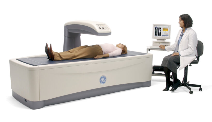
One of the most significant advantages of the PSMA PET scan is that it is a non-invasive imaging technique. Patients do not need to undergo painful or invasive procedures, such as biopsies, to obtain critical information about the extent of their prostate cancer. Additionally, the PSMA PET scan uses a low dose of radioactive material that is safe for patients.

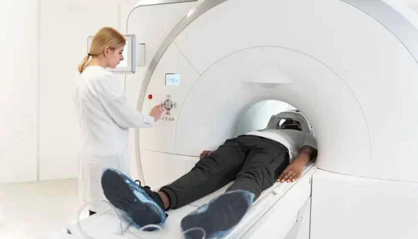
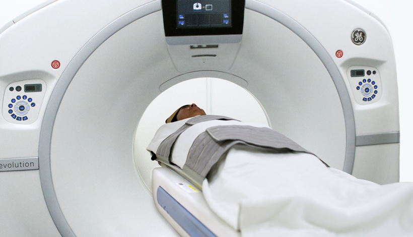
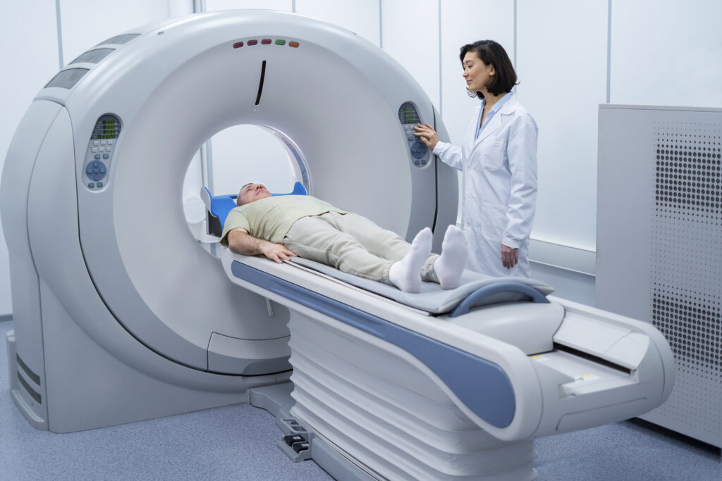
The procedure involves a small injection of a radiotracer, which binds to PSMA proteins on prostate cancer cells. The patient then lies on a table while a PET scanner captures detailed images of the body. The entire process typically takes 30 to 45 minutes and requires minimal preparation. The non-invasive nature of the procedure makes it a highly preferred option for prostate cancer patients, especially those who may already be dealing with the stress and discomfort of cancer treatment.

Pro Tip: At Ace Imaging, we ensure that all our patients are made comfortable throughout the process, and our team of experts closely monitors every step of the procedure.

4. Monitoring Treatment Response and Early Detection of Relapse

One of the biggest challenges in prostate cancer management is monitoring the effectiveness of treatments and detecting early relapses. The PSMA PET scan plays a crucial role in tracking how well the cancer is responding to treatment, whether it’s surgery, radiation therapy, or hormone therapy.

As prostate cancer responds to treatment, the PSMA PET scan can show whether the tumor is shrinking or whether new areas of metastasis are developing. This allows for timely adjustments to the treatment plan. In cases where prostate cancer is hormonally resistant or has become refractory to therapy, the PSMA PET scan can identify areas of recurrence before they show up on traditional imaging scans.

By catching cancer relapse early, doctors at Ace Imaging can implement targeted treatments sooner, improving the likelihood of a positive outcome. In some cases, this could mean offering more aggressive therapies or switching to different treatment options to control the disease.

5. Personalized Treatment Decisions and Better Patient Outcomes

With its high level of accuracy, the PSMA PET scan enables a more personalized treatment approach for prostate cancer patients. Armed with detailed information about the tumor’s exact location, spread, and response to treatments, doctors can make more informed decisions about how to manage the disease.

A more personalized approach to treatment allows patients to:

  • Avoid Over-Treatment: By accurately identifying the spread of cancer, doctors can avoid over-treating non-aggressive forms of prostate cancer, preserving patients’ quality of life.
  • Tailor Therapy Plans: Whether it’s surgery, radiation therapy, or systemic treatments like chemotherapy, doctors can customize the treatment plan based on the specific needs of the patient.
  • Improve Outcomes: By using the PSMA PET scan to monitor the progression of the disease and adapt treatment as needed, patients are more likely to achieve better outcomes and longer survival rates.

Conclusion

Prostate cancer can be a challenging diagnosis, but with advancements in imaging technology like the PSMA PET scan, patients in Thane and beyond now have access to cutting-edge diagnostic tools that can greatly improve the accuracy of their diagnosis and the effectiveness of their treatment. At Ace Imaging, we are proud to offer the PSMA PET scan in Mumbai as part of our comprehensive prostate cancer care services.

Whether you’re seeking a diagnosis, exploring treatment options, or monitoring the effectiveness of your current treatment, the PSMA PET scan provides crucial information that can guide your healthcare team in making the best decisions for your health.

PET Scans for Early Cancer Detection in Mumbai

The Benefits of PSMA PET Scans for Early Cancer Detection in Mumbai

Introduction In the fight against cancer, early and accurate diagnosis is crucial. The PSMA PET Scan in Mumbai has become a groundbreaking tool for detecting prostate cancer and other cancers at an early stage. This article delves into the benefits of this advanced imaging technique and how Ace Imaging in Mumbai is leading the way in offering this vital service.

What is a PSMA PET Scan? PSMA PET Scan (Prostate-Specific Membrane Antigen Positron Emission Tomography) is an advanced imaging test that provides detailed pictures of the prostate and other tissues where cancer may spread. This technology helps doctors identify cancer cells earlier and more accurately than traditional scans.

How PSMA PET Scan Works The PSMA Scan works by injecting a small amount of a radioactive tracer that binds to PSMA proteins commonly found on prostate cancer cells. This tracer makes the cancer cells glow on the PET scan, providing doctors with precise information about the location and extent of cancer.

Advantages of PSMA Scan for Early Cancer Detection 1. Enhanced Detection Capabilities The PSMA PET Scan in Mumbai stands out for its ability to detect even small cancerous lesions that other imaging techniques might miss. This is crucial for initiating early treatment.

  1. Accurate Staging and Treatment Planning For patients diagnosed with prostate cancer, knowing the exact stage is essential for effective treatment. Ace Imaging provides PSMA PET Scans that help map out the cancer’s reach, ensuring that doctors can create targeted treatment plans.
  2. Reduced Need for Multiple Tests Traditional imaging might require multiple tests to confirm cancer presence and spread. A PSMA Scan consolidates this process, offering comprehensive insights in a single session, saving both time and cost.

Why Choose PSMA Scan Over Other Imaging Techniques?

  • Higher Sensitivity and Specificity: The scan’s ability to identify cancerous tissues with greater sensitivity than CT or MRI scans.
  • Less Invasive: While being highly informative, a PSMA Scan is non-invasive and well-tolerated by patients.
  • Efficient Diagnosis: It provides quicker and more reliable results, which helps reduce the anxiety of waiting for multiple tests.

The Role of Ace Imaging in Delivering Quality PSMA PET Scans in Mumbai Ace Imaging in Mumbai is at the forefront of providing state-of-the-art diagnostic imaging services. Equipped with the latest technology and a team of skilled radiologists, Ace Imaging ensures that patients receive accurate, timely, and compassionate care.

What Sets Ace Imaging Apart?

  • Cutting-edge Technology: Utilizing the latest advancements in PET scan technology.
  • Experienced Team: Radiologists with specialized training in interpreting PSMA PET Scans.
  • Comprehensive Care: A patient-centered approach that emphasizes clear communication and tailored care plans.

Conclusion The PSMA PET Scan in Mumbai offered by Ace Imaging is revolutionizing early cancer detection. With unmatched precision, faster results, and enhanced detection capabilities, this advanced imaging technique is invaluable for patients and healthcare providers alike. Choosing Ace Imaging for a PSMA Scan ensures you receive the highest standard of diagnostic care, contributing to better outcomes and peace of mind.

LDCT scan in mumbai

Low dose CT : new technology for lung cancer screening – is it really worth?CTLow dose CT : new technology for lung cancer screening – is it really worth?

Low-dose computed tomography (LDCT) has gained attention as a screening tool for lung cancer, particularly in individuals at high risk for the disease. The use of LDCT for lung cancer screening has been studied extensively, and it has shown promise in detecting lung cancers at earlier, more treatable stages. 

Advantages of LDCT for Lung Cancer Screening:

Early Detection: LDCT has demonstrated a higher sensitivity in detecting small lung nodules compared to traditional chest X-rays. Early detection can lead to more effective treatment and improved outcomes.

Reduced Mortality: Clinical trials, such as the National Lung Screening Trial (NLST), have shown that lung cancer screening with LDCT can lead to a reduction in lung cancer mortality compared to chest X-ray screening.

Risk Stratification: LDCT is generally recommended for individuals at high risk for lung cancer, such as those with a significant smoking history. This helps in targeting the screening to those who are more likely to benefit.

Less Invasive than Biopsy: Detecting lung nodules early through LDCT may reduce the need for more invasive diagnostic procedures like lung biopsies.

Improved Survival Rates: Early-stage lung cancers often have better prognosis and treatment options. LDCT screening may contribute to improved survival rates by identifying cancers before they advance.

Pitfalls: 

False Positives:  LDCT may lead to false-positive results, where nodules are detected that are not cancerous. This can lead to additional tests, potential anxiety, and unnecessary interventions.

Radiation Exposure: Although LDCT uses lower radiation doses compared to standard CT scans, it still involves exposure to ionizing radiation. The balance between benefits and potential harm from radiation should be considered.

Appropriate Patient Selection: Identifying individuals at high risk for lung cancer is crucial for the effectiveness of LDCT screening. Screening should be targeted toward those who are most likely to benefit.

Recommendations:

The U.S. Preventive Services Task Force (USPSTF) recommends annual LDCT screening for individuals aged 50-80 years who have a 20 pack-year smoking history and currently smoke or have quit within the past 15 years.

Shared decision-making between patients and healthcare providers is essential to discuss the potential benefits and risks of LDCT screening based on individual factors.

In conclusion, LDCT for lung cancer screening can be valuable, especially for high-risk individuals, as it has the potential to detect lung cancer at earlier stages and reduce mortality. However, the decision to undergo screening should be individualized, considering the specific risks and benefits for each person. 

DEXA

Decoding DEXA scans : Understanding bone density assessment

Dual-energy X-ray absorptiometry (DEXA) scans are a commonly used diagnostic tool to assess bone density and are particularly valuable in diagnosing and monitoring conditions such as osteoporosis. Let’s break down the key aspects of decoding DEXA scans and understanding bone density assessment:

Principle of DEXA: DEXA uses two X-ray beams, each with different energy levels, to measure the absorption of these beams by bone and soft tissue. The attenuation of the X-ray beams by bone is directly related to bone mineral density (BMD).

Bone Mineral Density (BMD):

BMD is a measure of the amount of mineral (mostly calcium and phosphorus) per unit volume in bone. DEXA measures BMD at various skeletal sites, commonly the spine, hip, and forearm.

T-score and Z-score:

T-score: This score compares an individual’s BMD with that of a healthy young adult of the same sex. It is used in diagnosing osteoporosis.

T-score between +1 and -1: Normal

T-score between -1 and -2.5: Osteopenia (low bone mass)

T-score -2.5 or lower: Osteoporosis

Z-score: This score compares BMD with the average BMD of individuals of the same age, sex, and ethnicity.

Z-score below -2: Lower than expected for age (may indicate secondary causes of bone loss)

Regions of Interest (ROI): DEXA scans typically assess specific regions, including the lumbar spine, femoral neck, and total hip. These regions are prone to fractures and are crucial for diagnosing osteoporosis.

Osteoporosis and Fracture Risk: Osteoporosis is characterized by low bone mass and structural deterioration, leading to an increased risk of fractures. DEXA scans help identify individuals at risk for fractures, allowing for early intervention to prevent bone loss and fractures.

Follow-up and Monitoring: DEXA scans are used for monitoring changes in bone density over time, evaluating the effectiveness of treatment, and adjusting therapeutic approaches accordingly.

Factors Influencing BMD: Various factors, including age, sex, genetics, hormonal status, and lifestyle factors (nutrition, physical activity, smoking), influence BMD.

Clinical Recommendations: DEXA screening is recommended for postmenopausal women and older men with risk factors for fractures. The frequency of DEXA scans depends on individual risk factors and initial findings. Understanding DEXA scan results requires collaboration between patients and healthcare providers to interpret the findings in the context of overall health and risk factors. Regular assessments help in early detection and effective management of bone-related conditions.