Is CT scan done empty stomach

Is CT Scan Done Empty Stomach? Everything You Need to Know

Introduction

When patients are scheduled for a CT scan, one of the most common questions they ask is: “Is CT scan done empty stomach?” The answer depends on the type of CT scan being performed, whether contrast dye is used, and the area of the body being examined.

At Ace Imaging Centre, we believe patients feel more comfortable and confident when they are well-informed. This guide will explain in detail when fasting is necessary for a CT scan, what happens if you eat before the scan, how contrast material plays a role, and how to prepare for your appointment.

Is CT scan done empty stomach

What Is a CT Scan?

A CT (Computed Tomography) scan is a diagnostic imaging test that combines X-rays and computer technology to produce detailed cross-sectional images of the body. It helps doctors detect:

  • Tumors and cancers
  • Bone fractures
  • Blood vessel blockages
  • Internal injuries
  • Infections or inflammation
  • Neurological disorders

CT scans can be performed with or without contrast dye (oral, intravenous, or rectal), and preparation instructions may differ accordingly.

Is CT Scan Done Empty Stomach?

The requirement for fasting depends on the type of CT scan:

Type of CT ScanFasting Required?Reason
CT Scan without ContrastNoRegular CT scans without dye usually don’t need fasting.
CT Scan with IV ContrastYes (4–6 hours)Fasting reduces the risk of nausea or vomiting due to contrast injection.
CT Abdomen/Pelvis with Oral ContrastYes (6 hours)An empty stomach ensures better absorption and clearer images.
CT AngiographyYes (4–6 hours)Helps improve clarity of blood vessel imaging.
Emergency CT ScanDepends on situationIn emergencies, CT can be done without fasting if life-threatening.

Answer:
Yes, some CT scans need to be done on an empty stomach, especially those involving contrast dye. However, many routine scans can be performed without fasting.

Why Is Fasting Required for a CT Scan?

Doctors may ask you to fast before a CT scan for the following reasons:

  1. Reduce Risk of Nausea:
    Contrast dye can sometimes cause nausea. Having food in the stomach increases the risk of vomiting during or after the scan.
  2. Prevent Aspiration:
    If vomiting occurs, fasting lowers the risk of inhaling stomach contents into the lungs.
  3. Improve Image Clarity:
    An empty stomach, especially in abdominal scans, ensures that the stomach and intestines do not interfere with the visualization of organs.
  4. Better Contrast Absorption:
    Oral contrast works best when the stomach and intestines are empty, giving radiologists clear images.

What Happens If You Eat Before a CT Scan?

If you mistakenly eat before a scan where fasting is required, it may lead to:

  • Rescheduling of the scan
  • Inconclusive or unclear images
  • Increased risk of nausea or vomiting after contrast injection

Tip: Always inform your radiology team if you have eaten before the test. In some cases, the scan can still proceed if the situation is urgent.

How Many Hours of Fasting Are Required?

The standard fasting time before a CT scan with contrast is:

  • 4–6 hours before the procedure
  • Only small sips of water are usually allowed
  • Diabetic patients may receive special instructions regarding their medications and insulin

CT Scan Preparation: Step-by-Step

Here’s what you can expect when preparing for a CT scan:

1. Before the Scan

  • Follow your doctor’s instructions on fasting.
  • Remove metallic objects (jewelry, watches, belts).
  • Inform your doctor about:
    • Allergies to contrast dye
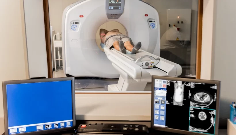
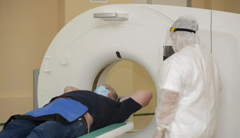
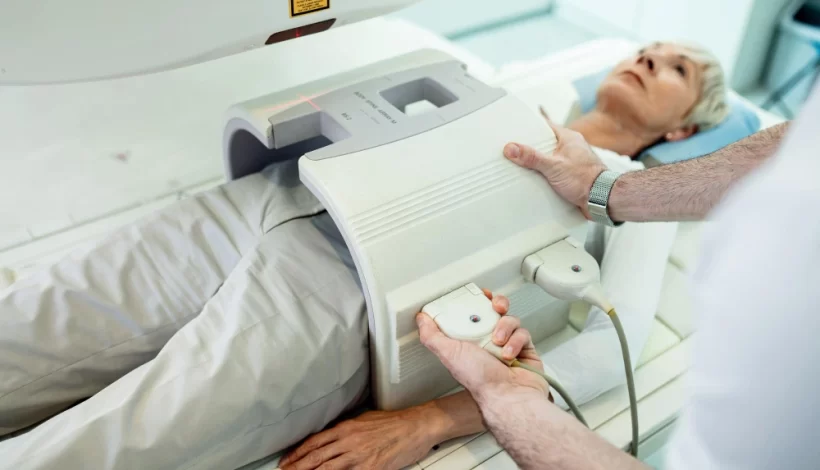
    • Pregnancy or possibility of pregnancy
    • Kidney disease or diabetes
    • Current medications

2. During the Scan

  • You will lie on a motorized table.
  • The table slides into the CT machine.
  • If contrast is used:
    • IV contrast may cause a warm sensation or metallic taste.
    • Oral contrast is usually given 1–2 hours before the scan.
  • The scan usually takes 15–30 minutes.

3. After the Scan

  • You may resume normal diet if no sedation is used.
  • Drink plenty of water to flush out contrast dye.
  • If you feel allergic reactions (rash, itching, shortness of breath), inform the radiologist immediately.

Special Considerations

1. For Diabetic Patients

  • Some medications (like metformin) may interact with contrast dye.
  • Patients are advised to stop metformin 48 hours after contrast scans.

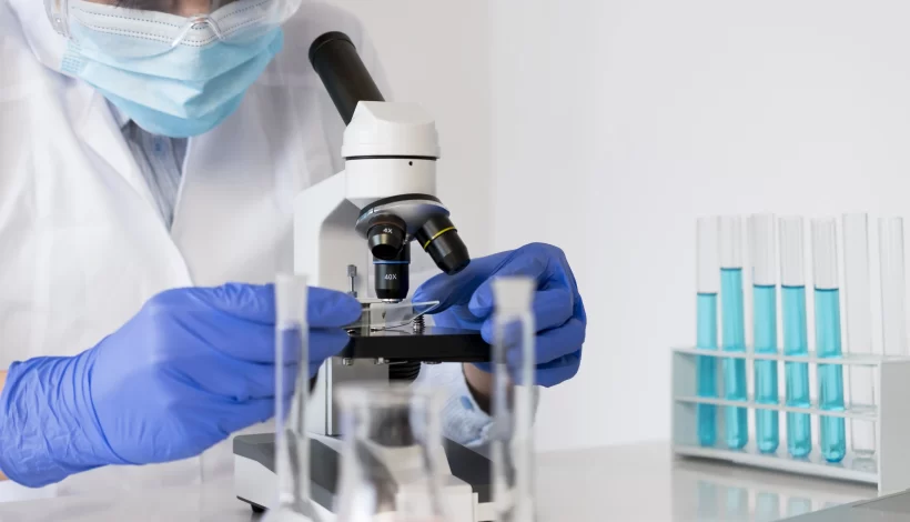
2. For Kidney Patients

  • Contrast dye may stress the kidneys. Blood tests (serum creatinine) may be required before the scan.

3. For Pregnant Women

  • CT scans involve radiation. Unless absolutely necessary, alternatives like ultrasound or MRI may be recommended.

4. For Children

  • Children may need fasting, depending on the type of CT scan.
  • In some cases, sedation is given to help them stay still.

Frequently Asked Questions (FAQs)

1. Is CT scan done empty stomach always?

No. Only CT scans involving contrast dye usually require fasting. Routine scans without contrast can often be done without fasting.

2. Can I drink water before a CT scan?

Yes. In most cases, small sips of water are allowed even during fasting.

3. How long should I fast before a CT scan?

Usually 4–6 hours. Your radiology team will give exact instructions.

4. What foods should I avoid before a CT scan?

Avoid solid foods, milk, coffee, or juice. Only clear fluids like water may be allowed.

5. What happens if I don’t fast before a contrast CT scan?

The scan may need to be rescheduled, or the images may not be clear enough for accurate diagnosis.

Tips for a Smooth CT Scan Experience

  • Arrive early to complete paperwork and preparation.
  • Wear loose clothing for comfort.
  • Bring medical reports and past imaging results.
  • Stay calm and relaxed—the procedure is painless and quick.
  • Hydrate well after the scan to flush out contrast dye.

Key Takeaways

  • Is CT scan done empty stomach?
    • Without contrast: Usually no fasting needed.
    • With contrast (oral or IV): Fasting 4–6 hours is required.
  • Always follow your doctor’s instructions for the specific type of scan you are undergoing.
  • Fasting ensures better safety, clearer imaging, and reduced complications.
  • If in doubt, contact your radiology centre before the scan.

Why Choose Ace Imaging Centre?

At Ace Imaging Centre, we provide:

  • State-of-the-art CT scan technology for accurate diagnosis
  • Patient-friendly guidance on fasting and preparation
  • Experienced radiologists and technicians ensuring safety and comfort
  • 24/7 support for emergency cases

We ensure every patient feels confident and informed before their scan. If you have questions like “Is CT scan done empty stomach?”, our expert team is here to guide you.

Conclusion

The answer to “Is CT scan done empty stomach?” depends largely on whether contrast dye is involved. For scans without contrast, fasting may not be necessary. However, for abdominal scans, angiography, or any test involving contrast dye, fasting for 4–6 hours is usually required.

At Ace Imaging Centre, we recommend that patients always follow their doctor’s preparation instructions for best results. Being informed about your scan helps reduce anxiety and ensures accurate, safe imaging.

    Book an appointment

    Fill form and get discount. Check Offers

    Top 5 Myths About MRI Scans

    Top 5 Myths About MRI Scans: Debunking Common Misconceptions

    Introduction

    Magnetic Resonance Imaging (MRI) is one of the most advanced diagnostic imaging tools used in modern medicine. Despite its accuracy and safety, many patients still feel anxious about undergoing an MRI scan due to widespread myths and misconceptions. These myths often prevent people from seeking timely medical help, leading to unnecessary delays in diagnosis and treatment.

    In this blog, we will debunk the Top 5 Myths About MRI Scans with clear explanations to help patients feel confident and informed before their procedure.

    What is an MRI Scan?

    An MRI scan uses a powerful magnetic field, radio waves, and a computer to create detailed images of the inside of the body. Unlike X-rays and CT scans, MRI does not use ionizing radiation, making it a safe and effective tool for diagnosing a wide range of conditions.

    MRI is commonly used for:

    • Brain and spinal cord imaging
    • Joint, ligament, and soft tissue evaluation
    • Detecting tumors and cancers
    • Heart and blood vessel imaging
    • Abdominal and pelvic organ scans

    Now, let’s break down the Top 5 Myths About MRI Scans and separate fact from fiction.

    Myth 1: MRI Scans Are Unsafe Because of Radiation

    One of the most common misconceptions is that MRI scans expose patients to harmful radiation, similar to X-rays or CT scans.

    The Truth:

    • MRI does not use radiation at all. Instead, it relies on magnetic fields and radiofrequency waves.
    • The magnetic field temporarily realigns hydrogen atoms in the body. When these atoms return to their normal position, they emit signals that are captured to create images.
    • Unlike CT scans, which use a small dose of ionizing radiation, MRI is completely free of radiation exposure.

    Why This Matters:

    This makes MRI scans particularly safe for patients of all ages, including children and pregnant women (with certain precautions).

    Myth 2: MRI Scans Are Only for Serious Conditions

    Some patients believe that an MRI scan is only prescribed for severe illnesses like cancer or brain tumors.

    The Truth:

    • MRI scans are versatile and can be used for both serious and routine medical evaluations.
    • Doctors often recommend MRI for:
      • Back pain and slipped disc
      • Sports injuries (ligaments, tendons, cartilage)
      • Chronic headaches or migraines
      • Knee or shoulder injuries
      • Abdominal pain and pelvic conditions

    Why This Matters:

    Even if you are not dealing with a life-threatening condition, your doctor may order an MRI scan to get a clearer view of soft tissues, which cannot be seen as well with X-rays or ultrasounds.

    Myth 3: MRI Scans Are Painful and Dangerous

    Many patients avoid MRI scans because they fear it might hurt or cause side effects.

    The Truth:

    • An MRI scan is completely painless. You won’t feel the magnetic field or radio waves.
    • The only discomfort comes from:
      • Staying still during the scan (usually 15–60 minutes)
      • Loud noises from the machine (earplugs or headphones are provided)
    • MRI is non-invasive and does not cause long-term side effects.

    Safety Considerations:

    • Patients with certain implants (such as pacemakers, cochlear implants, or metal fragments) must inform their doctor beforehand, as the magnetic field may interfere.
    • Otherwise, MRI is one of the safest diagnostic imaging procedures available.

    Myth 4: People with Claustrophobia Cannot Have an MRI

    Claustrophobia, or fear of enclosed spaces, is a common concern when it comes to MRI scans. Some patients believe they cannot undergo MRI if they have anxiety in confined spaces.

    The Truth:

    • While MRI scanners are enclosed tubes, there are solutions for patients with claustrophobia:
      • Open MRI machines: Wider and less enclosed, designed for comfort.
      • Sedation options: Mild sedatives may be used to help patients relax.
      • Music or communication systems: Many centers provide headphones with music or allow constant communication with the technologist.

    Why This Matters:

    Modern MRI technology is patient-friendly. If you are claustrophobic, you can still undergo MRI safely with the right support.

    Myth 5: MRI Scans Take Too Long and Are Inconvenient

    Some people think an MRI scan requires hours of preparation and scanning, making it too inconvenient for their schedule.

    The Truth:

    • A typical MRI scan takes 15 to 60 minutes, depending on the body part being imaged.
    • Preparation is minimal:
      • Remove metallic objects (jewelry, watches, belts).
      • Change into a hospital gown if required.
      • Answer safety questions about implants or medical devices.
    • Patients can usually return to normal activities immediately after the scan.

    Why This Matters:

    MRI scans are quick, efficient, and non-invasive. They provide valuable diagnostic information that can save time in long-term treatment planning.

    Summary Table: Myths vs. Facts About MRI Scans

    MythFact
    MRI uses harmful radiationMRI uses magnetic fields, not radiation
    MRI is only for serious diseasesMRI is used for both routine and critical conditions
    MRI is painful and riskyMRI is safe, painless, and non-invasive
    Claustrophobic patients cannot undergo MRIOptions like open MRI and sedation are available
    MRI takes too long and is inconvenientMost MRI scans take only 15–60 minutes

    Benefits of MRI Scans Over Other Imaging Techniques

    To better understand why MRI is often recommended, here’s a quick comparison:

    Imaging MethodRadiation ExposureBest ForLimitations
    X-rayYesBones, fracturesLimited soft tissue detail
    CT ScanYes (low dose)Trauma, chest/abdominal imagingRadiation exposure
    UltrasoundNoPregnancy, abdominal organsLimited in deep tissues
    MRINoBrain, spine, joints, soft tissues, tumorsLonger scan time, not suitable for patients with certain implants

    Tips for Patients Before an MRI Scan

    To make your MRI experience smoother:

    • Inform your doctor if you have implants, pacemakers, or metal fragments.
    • Avoid wearing metallic objects (jewelry, zippers, belts).
    • Stay relaxed – MRI scans are safe and painless.
    • Ask about sedation or open MRI if you have claustrophobia.
    • Follow any fasting instructions if contrast dye is required.

    Why Choose Ace Imaging Centre for MRI Scans?

    At Ace Imaging Centre, we understand that patients may feel anxious about MRI scans due to common myths and misconceptions. Our facility is designed to ensure:

    • State-of-the-art MRI technology for accurate results
    • Patient comfort with open MRI and relaxation options
    • Experienced radiologists and technicians who guide you at every step
    • Quick reporting and diagnosis to help your doctor plan treatment faster

    When you choose Ace Imaging Centre, you are choosing safety, accuracy, and care.

    Final Thoughts

    MRI scans are one of the safest and most reliable diagnostic tools in modern medicine. Unfortunately, myths about MRI scans often create unnecessary fear among patients. By understanding the facts, you can approach your MRI appointment with confidence.

    Remember:

    • MRI does not use radiation.
    • It is safe, painless, and effective.
    • Claustrophobia and convenience concerns can be easily managed.

    At Ace Imaging Centre, we are committed to making your MRI experience stress-free and accurate.

      Book an appointment

      Fill form and get discount. Check Offers

      DOTA Scan vs PET Scan

      DOTA Scan vs PET Scan – Complete Guide for Patients

      Introduction

      Medical imaging has revolutionised the way doctors diagnose and treat diseases. Two commonly discussed scans are the DOTA scan and the PET scan. While both involve advanced nuclear medicine techniques, they are not the same. Understanding the difference between a DOTA scan vs PET scan can help patients feel informed and confident before undergoing either procedure.

      This guide explains in detail what each scan is, how they work, when they are recommended, and their benefits, so you can make informed healthcare decisions with your doctor.

      What Is a PET Scan?

      PET scan stands for Positron Emission Tomography scan. It is an advanced imaging test that uses a small amount of radioactive tracer to detect abnormalities in the body at the cellular level.

      • Purpose: Detect metabolic changes in tissues before structural changes occur.
      • Common Uses:
        • Cancer detection, staging, and treatment monitoring
        • Heart disease evaluation
        • Brain disorders (e.g., Alzheimer’s, epilepsy)
      • Tracer Used: Commonly FDG (Fluorodeoxyglucose)

      How It Works:

      1. A radioactive tracer is injected into the bloodstream.
      2. The tracer travels to target organs or cells.
      3. The PET scanner detects gamma rays emitted from the tracer, creating detailed images of body function.

      What Is a DOTA Scan?

      A DOTA scan is a specialised type of PET scan used primarily to detect neuroendocrine tumours (NETs).

      • Full Form: DOTA refers to DOTATATE or DOTATOC, which are molecules that bind to somatostatin receptors on certain tumour cells.
      • Purpose: Specifically targets tumours with somatostatin receptors.
      • Tracer Used: Gallium-68 DOTATATE or Gallium-68 DOTATOC.

      How It Works:

      1. A radiolabelled DOTA compound is injected into the patient.
      2. It binds to somatostatin receptors present on certain tumours.
      3. PET/CT scanning captures high-contrast images showing tumour locations and spread.

      DOTA Scan vs PET Scan – Key Differences

      While both scans use PET technology, there are important differences in the tracer used, the target, and the clinical applications.

      FeaturePET Scan (FDG)DOTA Scan (Ga-68 DOTATATE)
      Main PurposeDetects abnormal metabolism in various tissuesDetects neuroendocrine tumours with somatostatin receptors
      Tracer UsedFDG (fluorodeoxyglucose)Ga-68 DOTATATE or DOTATOC
      Best ForCancer, brain, heart diseasesNeuroendocrine tumours (NETs)
      SpecificityGeneral metabolic imagingHighly specific to NETs
      PreparationFasting 4–6 hours beforeSimilar fasting; sometimes additional instructions for NET patients
      Scan Time20–45 minutes20–45 minutes

      When Doctors Recommend a PET Scan

      A PET scan may be recommended for:

      • Detecting cancer early
      • Determining the extent (stage) of cancer
      • Checking if treatment is working
      • Evaluating brain disorders like Alzheimer’s
      • Assessing heart muscle damage after a heart attack

      When Doctors Recommend a DOTA Scan

      A DOTA scan is usually ordered for:

      • Diagnosing suspected neuroendocrine tumours
      • Determining tumour spread (metastasis)
      • Evaluating treatment response in NETs
      • Locating small or hidden NETs not seen on other scans

      Procedure – What Patients Can Expect

      PET Scan Procedure

      1. Fasting for 4–6 hours.
      2. Injection of FDG tracer.
      3. Rest period to allow tracer distribution.
      4. Lying still inside the PET/CT scanner while images are taken.

      DOTA Scan Procedure

      1. Similar fasting guidelines.
      2. Injection of Ga-68 DOTATATE tracer.
      3. Waiting period (usually 45–60 minutes) for tracer uptake.
      4. PET/CT scanning.

      Advantages of PET Scan

      • Detects disease at an early stage.
      • Can assess how well treatment is working.
      • Useful in multiple medical fields (oncology, neurology, cardiology).

      Advantages of DOTA Scan

      • High accuracy for detecting NETs.
      • Can find tumours too small for conventional imaging.
      • Guides targeted therapy decisions.

      Limitations & Risks

      PET Scan Limitations:

      • False positives if inflammation is present.
      • Limited use for tumours with low glucose metabolism.

      DOTA Scan Limitations:

      • Only useful for tumours with somatostatin receptors.
      • Not for all cancer types.

      Risks for Both:

      • Small exposure to radiation.
      • Rare allergic reactions to tracer.
      • Mild discomfort at injection site.

      Preparation Tips for Patients

      • Follow fasting instructions carefully.
      • Avoid strenuous exercise 24 hours before the scan.
      • Inform your doctor about all medications and supplements.
      • For diabetic patients: Special scheduling may be required.

      DOTA Scan vs PET Scan – Choosing the Right Test

      The decision depends on:

      • Type of disease suspected
      • Tumour biology
      • Doctor’s assessment based on medical history and prior tests

      Example:
      If a doctor suspects lung cancer, a standard FDG PET scan may be chosen.
      If a patient has symptoms suggestive of a neuroendocrine tumour, a DOTA scan is more accurate.

      Cost & Availability

      • PET Scan: More widely available; cost varies by hospital and region.
      • DOTA Scan: Available in specialised imaging centres like Ace Imaging Centre; cost may be higher due to tracer specificity.

      Why Choose Ace Imaging Centre for PET or DOTA Scans?

      At Ace Imaging Centre, patients benefit from:

      • State-of-the-art PET/CT scanners
      • Experienced nuclear medicine specialists
      • Accurate diagnosis for better treatment planning
      • Comfortable and patient-friendly environment

      Our team ensures every scan is performed with utmost care, accuracy, and safety.

      Frequently Asked Questions

      Q1: Is a DOTA scan the same as a PET scan?
      No. A DOTA scan is a type of PET scan using a specific tracer for neuroendocrine tumours.

      Q2: Which is more accurate – DOTA scan or PET scan?
      Accuracy depends on the disease. For NETs, DOTA scans are superior. For general cancers, FDG PET scans are preferred.

      Q3: Is the radiation exposure dangerous?
      Both involve low radiation doses, generally considered safe under medical supervision.

      Q4: Can I eat before the scan?
      Fasting for 4–6 hours is usually required for both scans.

      Q5: How long do results take?
      Results are typically available within 24–48 hours.

      Conclusion

      While both scans use PET imaging technology, the DOTA scan vs PET scan debate is really about the right tool for the right job. A standard PET scan is versatile and used across many diseases, while a DOTA scan is highly specific for neuroendocrine tumours.

      At Ace Imaging Centre, we ensure patients get the correct scan based on their unique condition, leading to more accurate diagnoses and better treatment outcomes.

        Book an appointment

        Fill form and get discount. Check Offers

        Introduction to Pathology

        Introduction to Pathology: What It Is, How It Works, and Why It Matters

        Introduction to Pathology

        Pathology is the cornerstone of modern medicine. It forms the scientific basis for understanding diseases, diagnosing health conditions, and guiding clinical treatments. At Ace Imaging Center, we believe that every accurate diagnosis begins with high-quality pathology services. Whether it’s analyzing blood, tissue, or body fluids, pathology provides vital insights into the causes and progression of diseases.

        This blog serves as a comprehensive introduction to pathology, exploring its definition, branches, diagnostic methods, and its crucial role in patient care.

        What is Pathology?

        Pathology is the study of disease its causes, development, and effects on the body. It involves the examination of organs, tissues, bodily fluids, and cells to understand the nature of diseases and guide treatment decisions.

        Key Functions of Pathology:

        • Diagnose diseases like infections, cancer, or autoimmune conditions
        • Monitor the progression of chronic illnesses
        • Support preventive healthcare through early detection
        • Guide treatment plans and assess response to therapies

        What is a Pathology Test?

        A pathology test refers to any laboratory analysis performed on bodily samples—such as blood, urine, stool, tissues, or swabs—to detect abnormalities, monitor health, and diagnose diseases.

        Common Purposes of Pathology Tests:

        • Identifying infections (bacterial, viral, fungal)
        • Checking organ function (liver, kidney, thyroid)
        • Detecting cancerous cells
        • Measuring blood sugar or cholesterol levels
        • Monitoring medication effects

        Common Types of Pathology Tests:

        Test TypeExamplesPurpose
        Blood TestsCBC, LFT, KFT, Lipid ProfileGeneral health, organ function
        Urine TestsRoutine Urinalysis, CultureKidney function, infections
        Stool TestsOccult Blood, ParasitesDigestive health, GI bleeding
        BiopsiesBreast, liver, prostate tissue analysisCancer diagnosis
        CytologyPap smear, FNACDetecting cancer or abnormal cells
        MicrobiologySputum, blood, urine culturesIdentify infectious agents
        Molecular TestsPCR, genetic mutation panelsDNA/RNA analysis for precise diagnosis

        At Ace Imaging Center, all tests are performed using modern equipment and interpreted by expert pathologists to ensure accuracy.

        What is a Pathology Lab?

        A pathology lab (also called a diagnostic laboratory) is a specialized medical facility where tests are conducted on clinical specimens to obtain information about a patient’s health.

        Services Offered by a Pathology Lab:

        • Sample collection and preparation
        • Laboratory testing and analysis
        • Result interpretation by pathologists
        • Diagnostic support to clinicians

        Key Staff in a Pathology Lab:

        • Pathologists: Medical doctors who interpret test results
        • Lab Technicians: Perform testing procedures and maintain equipment
        • Phlebotomists: Collect blood and other samples
        • Microbiologists: Analyze infections
        • Molecular Biologists: Conduct advanced DNA-based testing

        At Ace Imaging Center, our NABL-accredited lab ensures safe, hygienic, and efficient testing with digital reporting and expert consultation.

        Branches of Pathology

        Pathology is a diverse field, divided into several branches based on the type of investigation and sample material. Each branch plays a specific role in medical diagnosis and research.

        BranchFocus Area
        Anatomical PathologyExamines tissues and organs for disease (e.g., biopsies, autopsies)
        Clinical PathologyAnalyzes blood, urine, and other body fluids to detect disease
        HematologyStudies blood and blood-forming tissues (e.g., anemia, leukemia)
        HistopathologyMicroscopic analysis of tissue samples
        CytopathologyStudies individual cells (e.g., Pap smear, FNAC)
        MicrobiologyIdentifies infections caused by bacteria, viruses, fungi, and parasites
        ImmunologyInvestigates immune responses and autoimmune diseases
        Molecular PathologyExplores genetic and molecular alterations in cells, especially in cancer diagnosis

        Each of these specialties is crucial in delivering comprehensive and accurate diagnostic services.

        Importance of Pathology in Modern Healthcare

        Pathology plays an indispensable role in diagnosing, managing, and preventing diseases. Without it, clinicians would be operating without a clear understanding of the patient’s condition.

        Why Pathology Matters:

        • Early Diagnosis: Detects diseases in their earliest stages, improving treatment outcomes.
        • Treatment Planning: Helps doctors choose the most effective treatments based on lab reports.
        • Prognosis Estimation: Predicts the likely course and outcome of a disease.
        • Monitoring Progress: Tracks changes in disease progression or response to treatment.
        • Screening: Identifies individuals at risk for specific conditions (e.g., cancer screening).

        Types of Pathology Tests

        There are numerous tests conducted under the field of pathology. These tests are typically categorized based on the sample type and the condition being investigated.

        Common Pathology Test Categories:

        1. Blood Tests
          • CBC (Complete Blood Count)
          • Blood Sugar Levels
          • Liver and Kidney Function Tests
          • Lipid Profile
          • Thyroid Function Tests
        2. Urine Tests
          • Routine Urinalysis
          • Urine Culture
          • Microalbumin Test
        3. Stool Tests
          • Occult Blood
          • Parasite Detection
        4. Biopsy and Histopathology
          • Tissue examination for cancer and other disorders
        5. Cytology
          • Pap smear
          • Fine Needle Aspiration Cytology (FNAC)
        6. Microbiological Culture Tests
          • Blood culture
          • Sputum culture
          • Wound swab culture
        7. Molecular and Genetic Testing
          • PCR tests
          • Cancer gene panels
          • BRCA testing for breast cancer

        Each test is performed with the highest standards of accuracy and reliability at Ace Imaging Center.

        The Pathology Testing Process: From Sample Collection to Report

        At Ace Imaging Center, the pathology process is handled with utmost precision to ensure accurate and timely results. Here’s a step by step breakdown of how it works:

        1. Sample Collection

        • Blood, urine, stool, tissue, or other biological materials are collected by trained phlebotomists or clinicians.
        • Proper labeling and handling are critical to avoid contamination or mix-ups.

        2. Sample Transportation

        • Samples are safely transported to the lab under controlled conditions to preserve their integrity.

        3. Laboratory Analysis

        • Advanced machines and microscopes analyze the samples based on the test ordered.
        • Pathologists, lab technicians, and medical microbiologists interpret findings.

        4. Result Interpretation

        • Qualified pathologists review results to confirm diagnoses.
        • The report includes findings, reference ranges, and comments for physicians.

        5. Report Delivery

        • Final reports are shared with the physician and patient digitally or in print.
        • Turnaround time depends on the complexity of the test.

        Who Performs Pathology Tests?

        Pathology tests are conducted by a team of highly trained healthcare professionals, each playing a vital role in ensuring diagnostic accuracy.

        Key Personnel in a Pathology Lab:

        • Pathologists: Doctors specialized in diagnosing disease through lab data and tissue analysis.
        • Lab Technicians: Skilled professionals who handle sample processing and test execution.
        • Microbiologists: Experts in detecting infections and antibiotic sensitivities.
        • Phlebotomists: Trained staff for collecting blood and other specimens from patients.
        • Molecular Biologists: Specialists in DNA/RNA testing and advanced cancer diagnostics.

        Role of Pathology in Cancer Diagnosis

        One of the most vital applications of pathology is in oncology (cancer diagnosis and treatment). Pathologists examine tissue biopsies to:

        • Confirm the presence of cancer
        • Identify cancer type (e.g., carcinoma, lymphoma)
        • Determine grade and stage
        • Guide oncologists in selecting appropriate therapies
        • Predict prognosis and risk of recurrence

        Example Tests in Cancer Pathology:

        TestPurpose
        Biopsy with HistopathologyConfirms type and aggressiveness of cancer
        Immunohistochemistry (IHC)Identifies specific tumor markers
        Molecular TestingDetects gene mutations (e.g., EGFR, KRAS)
        Liquid BiopsyNon-invasive detection of tumor DNA in blood

        Pathology and Preventive Healthcare

        Preventive medicine relies heavily on routine pathology screenings to detect issues before symptoms arise. This proactive approach helps in lowering disease burden and healthcare costs.

        Key Preventive Tests:

        • Annual health check-ups (CBC, blood sugar, lipids)
        • Cancer screenings (Pap smear, PSA test, mammography)
        • Thyroid and hormone levels
        • Diabetes markers (HbA1c)
        • Kidney function monitoring

        Digital Pathology: The Future of Diagnostics

        With the advent of technology, pathology is rapidly evolving. Digital pathology allows high-resolution images of tissue samples to be analyzed and shared remotely. This enhances:

        • Speed of diagnosis
        • Remote consultations (telepathology)
        • AI-assisted interpretation
        • Archival and tracking of patient data

        Ace Imaging Center is committed to adopting the latest advancements, ensuring world-class care.

        Why Choose Ace Imaging Center for Pathology Services?

        At Ace Imaging Center, we take pride in offering comprehensive pathology services backed by experienced professionals and advanced technologies.

        What Sets Us Apart?

        • NABL-accredited laboratory
        • State-of-the-art testing equipment
        • Rapid report turnaround
        • Expert team of certified pathologists and technicians
        • Hygienic and safe sample collection practices
        • Digital reports for easy access
        • Customizable health packages
        • Affordable rates with transparent billing

        We ensure that every report you receive is accurate, timely, and easy to understand because your health deserves nothing less.

        Frequently Asked Questions (FAQs)

        1. What is the difference between pathology and radiology?

        Pathology involves lab tests on tissues and body fluids, while radiology uses imaging like X-rays, CT scans, and MRIs to view internal body structures.

        2. How long does it take to get pathology test results?

        Basic tests may take a few hours, while complex ones like biopsies can take 2–5 days depending on the analysis needed.

        3. Are pathology tests painful?

        Most pathology tests are non-invasive or involve minor procedures like blood draws or swabs, which cause minimal discomfort.

        4. Can I get my pathology report online?

        Yes, Ace Imaging Center provides secure digital reports accessible through email or our online portal.

        5. Do I need a doctor’s prescription for pathology tests?

        Some tests require a doctor’s referral, but preventive screenings or health packages can often be booked directly.

        Final Thoughts

        This introduction to pathology highlights just how central this field is to every stage of patient care from prevention to diagnosis and treatment. Whether you’re undergoing a routine blood test or a complex biopsy, pathology is the key to unlocking critical insights about your health.

        Choosing a reliable and experienced diagnostic partner like Ace Imaging Center ensures you get the care, attention, and accuracy you deserve. Our mission is to empower patients and physicians with reliable lab support that makes all the difference.

        Book Your Test Today at Ace Imaging Center
        For safe, accurate, and affordable pathology services in Mumbai, trust the experts at Ace Imaging Center. Call us or visit our website to schedule your appointment.

          Book an appointment

          Fill form and get discount. Check Offers