
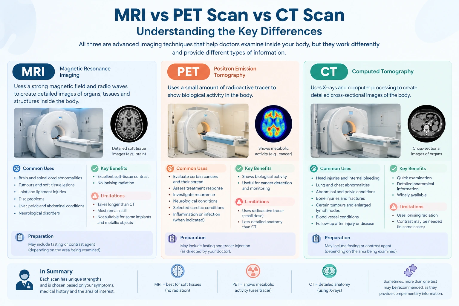
MRI, PET, and CT scans are medical imaging techniques that help doctors examine internal structures, investigate diseases, and guide treatment decisions. Although all three provide images of the body, they work differently and offer different types of medical information.
When comparing MRI vs PET scan vs CT scan, it is important to understand what each examination can reveal. MRI provides detailed images of soft tissues, CT produces cross-sectional images of internal structures using X-rays, and PET evaluates metabolic or other biological activity using a radioactive tracer.
Doctors select an imaging examination according to the patient’s symptoms, medical history, suspected condition, and the information required for diagnosis or treatment planning. In some situations, more than one imaging technique may be recommended because the tests provide complementary information.
This article explains the differences between MRI, PET, and CT scans, including their uses, preparation requirements, advantages, and limitations.
What Is an MRI Scan?
Magnetic Resonance Imaging (MRI) is a medical imaging technique that uses a strong magnetic field and radio waves to create detailed images of organs, tissues, and other structures inside the body.
Unlike CT scanning, MRI does not use ionising radiation. It is particularly useful for examining soft tissues, including the brain, spinal cord, muscles, ligaments, and certain internal organs.
Common Uses of MRI
Doctors may recommend an MRI scan to investigate:
- Brain and spinal cord abnormalities.
- Certain tumours and soft-tissue lesions.
- Ligament, tendon, and joint injuries.
- Intervertebral disc problems.
- Selected liver, pelvic, and abdominal conditions.
- Certain neurological disorders.
- Changes in soft tissues that require detailed assessment.
The specific type of MRI depends on the area being examined and the clinical question. Some examinations use a contrast agent to improve the visibility of particular tissues or abnormalities.
Advantages and Limitations of MRI
MRI provides excellent soft-tissue contrast and does not involve ionising radiation. However, the examination usually takes longer than a CT scan, and patients must remain relatively still during imaging.
The strong magnetic field also means that certain implants and metallic objects require careful assessment before the examination. Patients should inform the imaging team about pacemakers, implants, previous surgeries, and any possibility of metallic fragments in the body.
What Is a PET Scan?
Positron Emission Tomography (PET) is a type of nuclear medicine imaging that evaluates biological activity within the body. It uses a radiotracer, which is a small amount of radioactive material administered according to the examination protocol.
One commonly used tracer is fluorodeoxyglucose (FDG), a glucose-like substance that helps demonstrate glucose metabolism in tissues.
After administration, the tracer travels through the body and accumulates according to the biological processes being assessed. The scanner detects signals from the tracer and generates images that show its distribution.
Common Uses of PET Scans
PET imaging may be used to:
- Evaluate certain cancers and their spread.
- Assess treatment response in selected cancers.
- Investigate suspected cancer recurrence.
- Examine certain neurological conditions.
- Evaluate selected cardiac conditions.
- Investigate specific inflammatory or infectious conditions when indicated.
PET findings show patterns of biological activity, not cancer alone. Infection, inflammation, and normal physiological processes can also cause increased tracer uptake.
What Is a PET-CT Scan?
A PET-CT scan combines PET imaging with Computed Tomography. The PET component provides information about metabolic activity, while the CT component shows the anatomical location of that activity.
This combination can help doctors assess whether an area of increased tracer uptake corresponds to a particular organ, lymph node, or other structure.
PET-CT is commonly used in oncology, although its suitability depends on the suspected disease and the clinical purpose of the examination.
What Is a CT Scan?
Computed Tomography (CT) uses X-rays and computer processing to create detailed cross-sectional images of the body.
Unlike a conventional X-ray, which produces a projection image, CT provides multiple images that can be reconstructed into views of internal structures from different angles.
CT is frequently used when doctors need to examine organs, bones, blood vessels, or internal injuries. It can also be used to investigate certain cancers and support treatment planning.
Common Uses of CT Scans
A CT scan may be recommended to investigate:
- Head injuries and suspected internal bleeding.
- Lung and chest abnormalities.
- Abdominal and pelvic conditions.
- Bone injuries and complex fractures.
- Certain tumours and enlarged lymph nodes.
- Selected blood vessel conditions.
- Changes in organs following injury or disease.
Some CT examinations use an iodine-based contrast agent to improve the visibility of blood vessels, organs, or other structures. Contrast is not required for every CT scan.
Advantages and Limitations of CT
CT examinations are generally quick and provide detailed anatomical information. This makes CT particularly useful in emergency settings and when rapid assessment is necessary.
However, CT uses ionising radiation. Contrast-enhanced examinations also require consideration of previous contrast reactions, kidney function when relevant, and other patient-specific factors.
The potential benefits and risks should be considered when selecting the appropriate examination.
MRI vs PET Scan vs CT Scan: Key Differences
The main difference between these imaging techniques is the type of information they provide.
| Feature | MRI scan | PET scan | CT scan |
| Full form | Magnetic Resonance Imaging | Positron Emission Tomography | Computed Tomography |
| Imaging method | Magnetic field and radio waves | Radioactive tracer and specialised detector | X-rays and computer processing |
| Primary information | Detailed anatomy and soft-tissue characteristics | Metabolic or other biological activity | Detailed cross-sectional anatomy |
| Ionising radiation | No | Yes, from the radiotracer | Yes |
| Common applications | Brain, spine, joints, soft tissues, selected organs | Selected cancers, treatment assessment, neurological and cardiac applications | Trauma, chest, abdomen, bones, and selected cancers |
| Contrast or tracer | Contrast may be used | Radiotracer is generally required | Contrast may be used |
| Typical examination time | Often around 20–60 minutes, depending on protocol | Usually longer because tracer uptake takes time | Often completed within minutes |
| Important consideration | Metal implants and magnetic-field safety | Tracer-specific preparation and interpretation | Radiation exposure and possible contrast-related risks |
These are general comparisons. Actual examination times, preparation requirements, and diagnostic usefulness vary according to the body part, protocol, and clinical indication.
MRI vs PET Scan vs CT Scan for Cancer
MRI, PET, and CT scans can all contribute to cancer assessment, but they answer different clinical questions.
MRI for Cancer Evaluation
MRI can provide detailed information about the location and extent of abnormalities in soft tissues. Depending on the type of cancer, it may help assess involvement of nearby structures and support treatment planning.
For example, MRI is frequently used in the evaluation of selected brain, spinal, pelvic, and musculoskeletal tumours.
PET-CT for Cancer Evaluation
PET-CT can provide information about metabolic activity and its anatomical location. In selected cancers, it may help assess disease distribution, treatment response, or suspected recurrence.
However, not every cancer demonstrates high uptake of a particular PET tracer. Small lesions and some tumours with low tracer uptake may be difficult to detect.
CT for Cancer Evaluation
CT can show the size, location, and anatomical relationships of tumours and may help assess lymph nodes, lungs, abdominal organs, and other structures.
It is commonly used in cancer diagnosis, staging, and follow-up when clinically appropriate.
Important: No single imaging test is suitable for every type of cancer. The choice depends on the suspected cancer, the clinical question, and the information needed by the treating team.
MRI vs PET Scan vs CT Scan for Brain Conditions
The preferred examination for a brain condition depends on the symptoms and the suspected cause.
MRI: MRI provides detailed images of brain tissue and is frequently used to investigate certain tumours, neurological disorders, inflammation, and abnormalities involving the brain or spinal cord.
CT: CT can rapidly identify certain urgent problems, including intracranial bleeding and skull fractures. It may be selected when immediate imaging is required.
PET: Certain PET examinations can assess brain metabolism or other biological processes. They may be used in selected neurological evaluations, depending on the tracer and clinical indication.
These tests are not interchangeable. For sudden neurological symptoms, the treating team determines which examination is appropriate based on the urgency and suspected condition.
Which Scan Is More Accurate: MRI, PET, or CT?
There is no universal answer to which scan is more accurate. Diagnostic performance depends on the disease being investigated, the body part examined, the imaging protocol, and the characteristics of the abnormality.
MRI may provide more useful detail for certain soft-tissue conditions, while CT may be more appropriate for rapid assessment of particular injuries. PET-CT may provide additional information when metabolic activity is important to the clinical question.
A scan can also produce inconclusive, false-positive, or false-negative findings. For example, inflammation may cause increased uptake on PET imaging, while some small lesions may not be clearly visible.
Doctors interpret imaging results alongside symptoms, physical examination findings, laboratory tests, previous images, and other investigations. Additional testing may be necessary when the findings do not adequately explain the clinical picture.
Radiation Exposure: MRI vs PET Scan vs CT Scan
Radiation exposure is another important difference between these imaging techniques.
MRI Radiation Exposure
MRI does not use ionising radiation. However, it involves a strong magnetic field, and the safety of implants and other metallic objects must be assessed before scanning.
PET Scan Radiation Exposure
PET imaging uses a radioactive tracer. The amount of radiation depends on the tracer, the administered activity, and the examination protocol.
When PET is combined with CT, the CT component also contributes to the total radiation exposure.
CT Scan Radiation Exposure
CT uses X-rays to produce cross-sectional images. Radiation exposure varies according to the body part, scan settings, and examination protocol.
Doctors consider the expected diagnostic benefit when recommending imaging that involves ionising radiation. Patients who are pregnant or may be pregnant should inform their healthcare team before the examination.
How to Prepare for MRI, PET, and CT Scans
Preparation requirements vary according to the examination and the patient’s medical circumstances.
| Scan type | General preparation considerations |
| MRI | Inform staff about implants, pacemakers, metallic objects, previous surgeries, and claustrophobia. Follow instructions regarding contrast and clothing. |
| PET or PET-CT | Follow tracer-specific fasting instructions, discuss diabetes and medications, and follow guidance regarding physical activity and hydration. |
| CT | Follow instructions regarding food, fluids, and contrast when applicable. Report previous contrast reactions, relevant medical conditions, and possible pregnancy. |
Patients should not independently stop medication or change prescribed doses before an examination.
For all three imaging techniques, it is helpful to bring relevant medical records, previous imaging reports, and the referral information provided by the treating doctor.
Frequently Asked Questions
1. What is the main difference between MRI, PET, and CT scans?
MRI uses magnetic fields and radio waves to produce detailed images, CT uses X-rays to show internal anatomy, and PET uses a radioactive tracer to evaluate biological activity. PET-CT combines metabolic and anatomical information.
2. Is MRI safer than a CT scan?
MRI does not use ionising radiation, whereas CT does. However, safety depends on individual circumstances, including the need for diagnostic information, MRI implant compatibility, and any contrast-related considerations. The appropriate examination should be selected according to clinical need.
3. Is a PET scan the same as a CT scan?
No. PET assesses biological activity using a radiotracer, while CT provides anatomical images using X-rays. A PET-CT examination combines the two techniques.
4. Can MRI, PET, and CT scans detect cancer?
All three techniques can contribute to cancer assessment in appropriate situations. Their usefulness depends on the cancer type, location, size, and clinical question. Imaging findings alone may not establish a diagnosis, and biopsy or other investigations may be required.
5. Which scan takes the least time?
CT scanning itself is often completed within minutes. MRI commonly takes longer, while PET examinations require additional time for tracer administration and uptake before image acquisition. Actual duration varies by protocol.
6. Can a patient need more than one type of scan?
Yes. Different imaging techniques may provide complementary information. A doctor may recommend more than one examination when the findings from a single test are insufficient to answer the clinical question.
7. Does every PET scan include a CT scan?
No. PET can be performed as a standalone examination, although PET-CT is widely used. The appropriate technique depends on the clinical indication and available protocol.
Conclusion
MRI, PET, and CT scans serve different purposes in medical imaging. MRI provides detailed soft-tissue images without ionising radiation, CT offers rapid and detailed anatomical assessment using X-rays, and PET evaluates biological activity using a radioactive tracer.
Understanding the differences between MRI vs PET scan vs CT scan can help patients understand why different examinations may be recommended. However, the choice should be based on the patient’s symptoms, medical history, suspected condition, and the diagnostic information required.
Patients should discuss questions about preparation, contrast agents, radiation exposure, and examination suitability with their treating doctor and imaging team.
Medical disclaimer: This article is intended for general educational purposes and does not replace professional medical advice, diagnosis, or treatment. The selection and interpretation of medical imaging examinations should be based on individual clinical circumstances and the advice of qualified healthcare professionals.
Name: Ace Imaging Center
Address: Bezolla Complex, 102, VN Purav Marg, Swastik Park, Chembur, Mumbai, Maharashtra 400071
contact: 081693 60334