
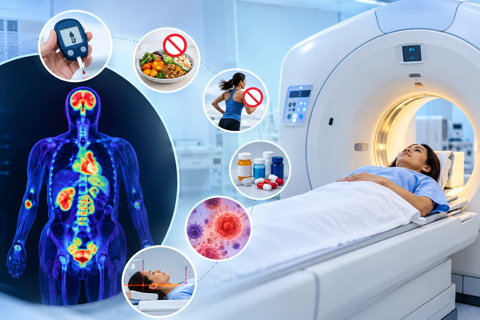
A Positron Emission Tomography (PET) scan is a medical imaging examination used to evaluate metabolic activity within the body. It can help doctors investigate certain cancers, assess treatment response, and evaluate selected neurological, cardiac, and inflammatory conditions. When combined with Computed Tomography (CT), a PET-CT scan provides functional and anatomical information that supports clinical assessment.
However, PET scan findings can be influenced by several factors, including blood glucose levels, food intake, physical activity, medications, inflammation, and the characteristics of the condition being investigated. Understanding these factors helps patients prepare appropriately and understand the limitations of the examination.
What can affect PET scan accuracy? The answer depends on the tracer used, the patient’s medical condition, the purpose of the examination, and the technical quality of the images. Although some factors can be managed through preparation, others are related to the biological characteristics of the disease or the limitations of medical imaging.
What Is a PET Scan and How Does It Work?
A PET scan uses a radiotracer, which is a small amount of a radioactive substance, to examine biological processes within the body. One commonly used tracer is fluorodeoxyglucose (FDG), a glucose-like substance that helps demonstrate how tissues use glucose.
After the tracer is injected into a vein, it travels through the bloodstream and accumulates in tissues according to their metabolic activity. The scanner detects signals from the tracer and creates images showing its distribution.
A PET-CT examination combines these functional images with CT images, helping doctors identify the anatomical location of areas showing altered metabolic activity.
PET scans may be used to:
- Investigate and stage certain cancers.
- Assess whether cancer has spread to other parts of the body.
- Evaluate metabolic changes during or after treatment.
- Investigate suspected recurrence of certain cancers.
- Assess selected neurological and cardiac conditions.
- Evaluate specific infections or inflammatory conditions when indicated.
Increased tracer uptake does not automatically indicate cancer, and a scan without an obvious abnormality does not exclude every disease. Results must be interpreted alongside the patient’s symptoms, medical history, physical examination, and other investigations.
What Can Affect PET Scan Accuracy?
Several patient-related, biological, and technical factors can influence PET scan findings. The importance of each factor varies according to the tracer and imaging protocol used.
1. Blood Glucose Levels
Blood glucose levels can influence the distribution of FDG during a PET scan. Because FDG behaves similarly to glucose, elevated blood glucose can compete with the tracer and alter its uptake in different tissues.
High glucose levels may affect image interpretation and the visibility of certain abnormalities. The degree of interference depends on the blood glucose level, the clinical indication, and the imaging protocol.
Patients with diabetes may require specific preparation instructions because blood glucose management and the timing of insulin administration can influence FDG distribution.
To prepare appropriately:
- Follow the fasting instructions provided by the imaging centre.
- Inform the medical team if you have diabetes.
- Share information about your diabetes medicines and usual glucose levels.
- Follow specific instructions about insulin and other glucose-lowering medications.
- Inform the staff if your blood glucose is unusually high or low.
Medication doses should not be changed independently. The treating doctor and imaging team can determine whether any adjustments or changes in scan timing are necessary.
2. Eating or Drinking Before the Examination
Food intake before an FDG PET scan may affect insulin levels and glucose metabolism. Eating shortly before the examination, particularly carbohydrate-rich foods, can alter tracer uptake in muscles and other tissues.
For many FDG PET scans, patients are instructed to fast for approximately four to six hours. However, fasting requirements depend on the tracer, examination protocol, and individual medical circumstances.
Plain water is generally permitted and often encouraged, unless different instructions have been provided.
Patients should confirm whether beverages such as tea, coffee, milk, juice, and other caloric drinks are permitted before their appointment.
Following the prescribed preparation instructions helps reduce avoidable changes in tracer distribution.
3. Recent Physical Activity
Physical activity increases glucose utilisation in working muscles. Since FDG uptake is influenced by glucose metabolism, strenuous exercise before a scan can lead to increased tracer accumulation in muscles.
This may make some areas more difficult to interpret or affect the appearance of surrounding tissues.
Patients are commonly advised to avoid strenuous exercise for approximately 24 hours before an FDG PET scan, or for the period specified by the imaging team.
Preparation may include:
- Avoiding vigorous workouts and heavy physical exertion.
- Following instructions about running, cycling, and other strenuous activities.
- Resting quietly during the tracer uptake period.
- Informing the staff about recent intense physical activity.
The recommended restrictions may vary according to the examination and the patient’s medical circumstances.
4. Medications and Recent Medical Treatments
Certain medications can influence tracer distribution or the appearance of tissues on PET images.
For example, insulin administered too close to an FDG PET scan may increase tracer uptake in muscles. Other medicines that affect glucose metabolism or inflammation may also influence findings, depending on the clinical situation.
Recent chemotherapy, radiotherapy, immunotherapy, surgery, or other medical procedures can cause changes in tissue metabolism. These changes may complicate the interpretation of findings when treatment effects resemble disease activity.
Patients should inform the imaging team about:
- Prescription and non-prescription medicines.
- Insulin and other diabetes treatments.
- Recent chemotherapy or radiotherapy.
- Immunotherapy and other cancer-related treatments.
- Recent surgery, biopsies, or invasive procedures.
- Recent changes in medication.
Patients should not stop prescribed medication without medical advice. The timing of the examination should be determined according to the clinical question and the relevant imaging protocol.
5. Infection and Inflammation
PET scans identify metabolic activity rather than cancer alone. Activated inflammatory cells can also accumulate FDG, producing areas of increased tracer uptake.
Infections, inflammatory disorders, and healing tissues may therefore produce findings that resemble malignancy.
For example, an inflamed lymph node may demonstrate increased uptake even when the cause is not cancer. Similarly, inflammation around a recent surgical site may affect the appearance of the region being examined.
The radiologist considers the pattern and location of uptake alongside the patient’s symptoms, medical history, laboratory results, and other imaging findings.
Patients should inform their doctor about recent infections, fever, inflammatory conditions, injuries, or surgical procedures before the examination.
6. Movement During Image Acquisition
Movement during a PET-CT scan can reduce image quality. It may cause blurring, misalignment between PET and CT images, or difficulty identifying the precise location of an abnormality.
Movement may occur because of discomfort, coughing, anxiety, involuntary muscle activity, or difficulty remaining in one position.
Patients can help minimise movement by following positioning instructions, wearing suitable clothing, and informing the staff about pain or physical limitations.
If movement significantly affects image quality, additional imaging may sometimes be necessary.
7. Timing Between Tracer Injection and Scanning
The interval between tracer administration and image acquisition is an important part of PET scan preparation.
After injection, the tracer requires time to distribute through the body. For many FDG PET scans, image acquisition begins approximately 60 minutes after injection, although the precise interval varies by protocol.
The timing affects tracer distribution and the appearance of tissues. Imaging outside the intended protocol may make comparisons with previous examinations more difficult.
The imaging team determines the appropriate uptake period based on the tracer, the clinical indication, and the examination protocol.
8. Size and Location of an Abnormality
The size and location of a lesion can influence whether it is visible on a PET scan.
Very small abnormalities may be difficult to detect because of the scanner’s spatial resolution. The partial-volume effect can also cause tracer uptake in small lesions to appear lower than it actually is.
Some tumours demonstrate relatively low FDG uptake, which may make them less conspicuous. Other lesions may be difficult to distinguish because of normal tracer activity in nearby tissues.
For example, normal physiological uptake in the brain, heart, kidneys, and urinary tract can affect the visibility of abnormalities in or around these structures.
A negative PET scan does not exclude every type of cancer or disease. Depending on the clinical circumstances, further imaging, laboratory testing, or biopsy may be necessary.
9. Body Characteristics and Image Quality
Body size and tissue characteristics can influence image noise and the visibility of smaller abnormalities.
In some patients, greater tissue thickness can reduce image clarity. Modern scanners and image reconstruction techniques help address these limitations, but they cannot eliminate every source of image noise.
Body composition and the distribution of muscle and fat may also influence tracer uptake.
These factors do not automatically mean that a scan is inaccurate. Instead, they are considered when assessing the quality of the images and interpreting the findings.
10. Scanner Performance and Imaging Protocol
PET scan interpretation depends partly on the technical quality of the images.
Factors that may influence image quality include:
- Scanner calibration and maintenance.
- Appropriate tracer administration.
- Image acquisition duration.
- Patient positioning.
- Attenuation correction and image reconstruction.
- Standardised imaging protocols.
- The availability of relevant previous scans.
Quality assurance procedures and appropriate technical protocols help reduce avoidable imaging errors. However, technical quality alone cannot overcome every biological limitation of a PET scan.
11. Medical History and Previous Imaging
Clinical information is important when interpreting PET scan findings.
Details about previous diagnoses, symptoms, treatments, surgery, infections, and other investigations can help the radiologist assess the significance of an abnormal area.
Previous imaging studies may also help determine whether a finding is new, unchanged, or has changed over time.
Patients should provide relevant medical records and inform the imaging team about their clinical history. The referring doctor and radiologist can use this information to interpret the results in context.
Factors That Can Affect PET Scan Accuracy: Summary
|
Factor |
Potential influence on findings |
|
Elevated blood glucose |
May alter FDG distribution and uptake |
|
Food intake |
May affect insulin levels and glucose metabolism |
|
Strenuous exercise |
May increase tracer uptake in muscles |
|
Medications |
May influence tracer distribution or tissue metabolism |
|
Infection or inflammation |
May cause increased uptake unrelated to cancer |
|
Recent surgery or treatment |
May produce healing-related or treatment-related changes |
|
Movement |
May cause image blurring or misalignment |
|
Small lesion size |
May reduce visibility because of spatial resolution |
|
Scanner performance |
May affect image clarity and detection |
|
Incomplete medical history |
May make findings more difficult to interpret in context |
How Should Patients Prepare for a PET Scan?
Preparation requirements depend on the tracer and examination being performed. Patients should follow the instructions provided by the imaging team rather than relying on general advice alone.
Common preparation considerations include:
- Fasting: Confirm when to stop eating and which drinks are permitted.
- Diabetes management: Obtain specific instructions regarding blood glucose monitoring and medication timing.
- Physical activity: Follow the recommended restrictions on strenuous exercise.
- Medication history: Share information about prescribed medicines and recent treatment.
- Medical history: Report recent infections, surgery, biopsies, or other relevant procedures.
- Hydration: Drink water as instructed.
- Clothing: Follow guidance regarding comfortable clothing and metal accessories.
- Previous reports: Bring relevant medical records and earlier imaging studies, when available.
- Pregnancy or breastfeeding: Inform the medical team beforehand, if applicable, so the examination can be assessed appropriately.
If any preparation instruction is unclear, patients should contact the imaging team before the examination.
Can a PET Scan Produce False-Positive or False-Negative Findings?
Yes. PET scans have diagnostic limitations and can produce false-positive or false-negative findings.
A false-positive finding occurs when an area appears suspicious even though the suspected disease is not present. Inflammation, infection, and healing after surgery may contribute to this type of finding.
A false-negative finding occurs when a disease or abnormality is present but is not detected by the scan. Small lesion size, low tracer uptake, and other imaging limitations may contribute.
The significance of a finding depends on the clinical context. Doctors may recommend additional imaging, laboratory tests, follow-up examinations, or biopsy when necessary.
PET scan results should not be used in isolation to establish or exclude every diagnosis. They are interpreted alongside other relevant clinical information.
When May a Repeat PET Scan Be Necessary?
A repeat PET scan may be considered when the original images are technically inadequate, preparation requirements were not met, or the examination did not sufficiently answer the clinical question.
Repeat imaging may also be used when clinically indicated to assess treatment response or investigate suspected disease recurrence.
However, an unexpected result does not automatically mean that another scan is required. The doctor may first review the original images, compare previous studies, or recommend a different investigation.
PET and PET-CT examinations involve ionising radiation from the radiotracer and, where applicable, the CT component. Repeat imaging should therefore be based on clinical need and an appropriate assessment of potential benefits and risks.
Frequently Asked Questions
1. What can affect PET scan accuracy the most?
For FDG PET scans, blood glucose levels, fasting, physical activity, medication timing, and the interval between tracer injection and image acquisition are important factors. Inflammation, lesion characteristics, and image quality can also influence interpretation.
2. Can high blood sugar affect PET scan results?
Yes. Elevated blood glucose can alter FDG uptake and distribution. Patients with diabetes should follow the specific preparation and medication instructions provided by their healthcare team.
3. Can eating before a PET scan affect the results?
Yes. Eating before an FDG PET scan may affect insulin levels and tracer distribution. Many protocols require fasting for several hours, but the exact instructions depend on the examination.
4. Can inflammation look like cancer on a PET scan?
Inflammation and certain infections can cause increased tracer uptake that resembles cancer. Additional clinical information or investigations may be required to determine the cause.
5. Can a PET scan miss cancer?
Yes. Some small lesions or tumours with low FDG uptake may not be clearly visible. A negative PET scan does not exclude every type of cancer, particularly when clinical suspicion remains.
6. Should I stop my medication before a PET scan?
Do not stop or change prescribed medication without medical guidance. Inform the imaging team about all medicines so that appropriate preparation instructions can be provided.
7. Is a PET-CT scan always accurate?
No diagnostic imaging examination is completely accurate. PET-CT provides functional and anatomical information, but its findings must be interpreted alongside the patient’s medical history and other relevant investigations.
Conclusion
PET scan accuracy depends on several factors, including blood glucose levels, fasting, physical activity, medications, inflammation, recent treatment, movement, lesion characteristics, and technical image quality.
Following the prescribed preparation instructions and providing accurate medical information can help reduce avoidable factors that affect image interpretation. Understanding the limitations of PET imaging is equally important, as neither increased nor decreased tracer uptake establishes a diagnosis on its own.
The findings should be interpreted by qualified medical professionals in the context of the patient’s symptoms, medical history, and other investigations.
Medical disclaimer: This article is intended for general educational purposes only. It does not replace individual medical advice, diagnosis, or treatment. PET scan preparation and interpretation may vary according to the tracer used, the clinical indication, and the patient’s medical condition. Patients should follow the instructions provided by their treating doctor and imaging team.
Name: Ace Imaging Center
Address: Bezolla Complex, 102, VN Purav Marg, Swastik Park, Chembur, Mumbai, Maharashtra 400071
contact: 081693 60334