
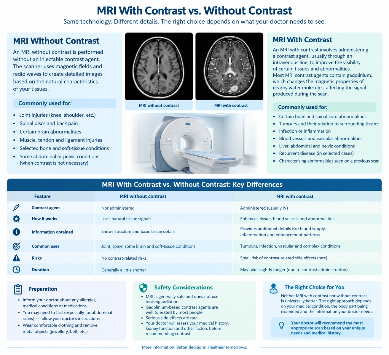
Magnetic Resonance Imaging (MRI) is a medical imaging technique that uses a strong magnetic field and radio waves to produce detailed images of organs, tissues, and other structures inside the body. It is commonly used to investigate neurological conditions, joint injuries, spinal problems, and abnormalities affecting various organs.
When an MRI is recommended, patients may be asked to undergo an examination with contrast or without contrast. The difference lies in whether a contrast agent is administered to improve the visibility of certain tissues, blood vessels, or abnormalities.o
Understanding MRI with contrast vs. without contrast can help patients know what to expect during the examination and why a particular approach may be recommended. However, neither method is universally better. The appropriate examination depends on the medical condition being investigated, the body part being examined, and the information required by the treating doctor.
This article explains how MRI contrast works, when each type of scan may be used, and the preparation and safety considerations patients should understand.
What Is an MRI Scan Without Contrast?
An MRI without contrast is performed without administering an injectable contrast agent. The scanner uses magnetic fields and radio waves to generate images based on the natural characteristics of tissues.
Different tissues produce different signal patterns, allowing radiologists to assess their structure and identify abnormalities. Multiple imaging sequences may be used to provide additional information about the area being examined.
Common Uses of MRI Without Contrast
An MRI without contrast may be recommended for:
- Assessing certain knee, shoulder, and other joint injuries.
- Examining spinal discs and investigating selected causes of back pain.
- Evaluating certain brain abnormalities.
- Investigating some muscle, tendon, and ligament injuries.
- Assessing selected bone and soft-tissue conditions.
- Examining particular abdominal or pelvic structures when contrast is not necessary.
The decision depends on the suspected condition and the examination protocol. Some conditions can be evaluated adequately without contrast, while others require additional information that contrast-enhanced imaging may provide.
Advantages of MRI Without Contrast
MRI without contrast offers several practical advantages:
- No intravenous contrast injection is required.
- It avoids risks specifically associated with the administered contrast agent.
- It can provide detailed images of many soft-tissue structures.
- It may be sufficient for the diagnostic question being investigated.
An MRI without contrast is not necessarily less informative. Its suitability depends on whether the images can adequately answer the clinical question.
What Is an MRI Scan With Contrast?
An MRI with contrast involves administering a contrast agent, usually through an intravenous line, to improve the visibility of certain tissues and abnormalities.
Most MRI contrast agents contain gadolinium, a metal used in a specially formulated compound. Following administration, the agent changes the magnetic properties of nearby water molecules, affecting the signal produced during MRI.
These changes can help radiologists assess tissue enhancement, blood supply, inflammation, and other characteristics of an abnormality.
Contrast is not required for every MRI examination. It is used when the additional information is expected to be clinically useful.
Common Uses of MRI With Contrast
An MRI with contrast may be recommended for:
- Evaluating certain brain and spinal cord abnormalities.
- Assessing selected tumours and their relationship with surrounding tissues.
- Investigating suspected infection or inflammation.
- Evaluating certain blood vessels and vascular abnormalities.
- Assessing some liver, abdominal, and pelvic conditions.
- Investigating suspected recurrent disease in selected clinical situations.
- Characterising abnormalities identified on an earlier examination.
For some conditions, doctors may request images both before and after contrast administration. Comparing the images can help identify changes that may not be fully characterised by non-contrast imaging alone.
How Does Contrast Improve MRI Images?
Contrast enhancement occurs when the administered agent changes the signal characteristics of tissues.
Different abnormalities may show different enhancement patterns. For example, certain tumours, inflammatory changes, and areas with altered blood-brain barrier integrity may enhance after contrast administration.
However, enhancement does not automatically indicate cancer or another specific disease. Benign conditions, inflammation, and normal tissue characteristics can also produce enhancement.
Radiologists interpret these patterns alongside other MRI sequences, the patient’s medical history, and relevant clinical findings.
MRI With Contrast vs. Without Contrast: Key Differences
The main difference between these examinations is whether a contrast agent is administered and whether the additional information it provides is needed.
|
Feature |
MRI without contrast |
MRI with contrast |
|
Contrast agent |
Not administered |
Usually administered intravenously |
|
Image information |
Shows tissue structure and signal characteristics |
Adds information about tissue enhancement and contrast distribution |
|
Common applications |
Selected joint, spine, brain, and soft-tissue examinations |
Selected tumour, inflammatory, vascular, and post-treatment evaluations |
|
Injection required |
Usually not for contrast |
Usually yes |
|
Additional safety considerations |
MRI magnetic-field safety still applies |
MRI safety plus assessment of contrast-related risks |
|
Kidney function assessment |
Usually not required solely for a non-contrast examination |
May be relevant depending on the contrast agent and the patient’s medical history |
|
Examination duration |
Depends on the protocol |
May take longer because additional sequences are acquired |
|
Diagnostic suitability |
Appropriate when contrast is unnecessary |
Appropriate when enhancement information is needed |
The exact examination protocol varies according to the body part, suspected condition, scanner, and clinical indication.
When Is MRI With Contrast Recommended?
Contrast-enhanced MRI is considered when information about tissue enhancement may help answer the clinical question.
1. Brain and Neurological Conditions
MRI with contrast may be used to evaluate selected brain tumours, infections, inflammatory conditions, and abnormalities affecting the brain or its coverings.
In some situations, contrast helps distinguish different types of tissue changes or assess the extent of an abnormality.
However, many brain MRI examinations can be performed without contrast, depending on the reason for imaging.
2. Tumour Evaluation
Contrast may help characterise certain tumours by showing how an abnormality enhances and how it relates to surrounding structures.
It can also be useful in selected follow-up examinations when doctors need to evaluate changes after treatment.
Enhancement patterns are not sufficient on their own to establish a diagnosis. The findings may need to be considered alongside other imaging studies, laboratory investigations, or biopsy results.
3. Infection and Inflammation
Certain infections and inflammatory conditions can cause changes in tissue enhancement.
Contrast-enhanced MRI may help assess the location and extent of an abnormality when clinically indicated.
The choice depends on the suspected condition and whether contrast is expected to provide additional diagnostic information.
4. Blood Vessels and Selected Organ Conditions
Some MRI examinations use contrast to assess blood vessels or evaluate abnormalities in organs such as the liver and pelvis.
The appropriate technique depends on the suspected disease and the type of information required. Some vascular MRI examinations can also be performed without contrast using specialised sequences.
When Is MRI Without Contrast Sufficient?
MRI without contrast may provide enough information when the examination is intended to assess structural changes that can be evaluated using standard MRI sequences.
Examples include:
- Many routine examinations of the knee and shoulder.
- Assessment of selected spinal disc abnormalities.
- Certain investigations of back pain.
- Evaluation of some joint and soft-tissue injuries.
- Selected brain examinations where contrast is not clinically necessary.
For example, an MRI performed to investigate a suspected ligament injury may not require contrast. However, the decision can change if the doctor is investigating infection, a tumour, or a different underlying condition.
The choice should be based on the clinical question rather than a general preference for one type of MRI.
Is MRI With Contrast Safe?
MRI contrast agents are generally well tolerated when used appropriately, but they are not completely free of risk.
Most gadolinium-based contrast agents have a good safety profile. Nevertheless, patients should inform the imaging team about relevant medical conditions and any previous reaction to an MRI contrast agent.
Possible Side Effects
Some patients may experience mild, temporary effects, including:
- Nausea.
- Headache.
- Dizziness.
- A feeling of coldness or discomfort during injection.
- Discomfort at the injection site.
Allergic-like reactions can occur, although severe reactions are uncommon.
A history of a previous reaction to a gadolinium-based contrast agent should be discussed before the examination. The imaging team can assess the circumstances and determine appropriate precautions.
Kidney Function and MRI Contrast
Kidney function is an important consideration for certain patients receiving gadolinium-based contrast agents.
In people with severe kidney impairment, some agents have been associated with a rare condition called nephrogenic systemic fibrosis. The risk depends on the specific agent and the patient’s medical circumstances.
Modern contrast agents differ in their safety profiles. The healthcare team determines whether kidney function assessment is necessary and which agent is appropriate.
Patients should inform the imaging team if they have kidney disease, are receiving dialysis, or have been advised that their kidney function is significantly reduced.
Pregnancy and Breastfeeding
If you are pregnant or may be pregnant, inform your doctor and the imaging team before an MRI examination.
Gadolinium-based contrast is generally avoided during pregnancy unless the expected clinical benefit justifies its use after an individual assessment.
For breastfeeding patients, routine interruption of breastfeeding is generally not necessary after administration of most gadolinium-based contrast agents. However, patients should discuss individual concerns with their healthcare professional.
How to Prepare for an MRI With or Without Contrast
Preparation depends on the body part being examined and the imaging protocol.
Before the Examination
Patients should:
- Inform the imaging team about pacemakers, implants, and previous surgeries.
- Report any previous reaction to an MRI contrast agent.
- Inform staff about kidney disease, pregnancy, or other relevant medical conditions.
- Follow instructions about eating and drinking.
- Ask whether any recent blood test or kidney function assessment is required.
- Remove metal objects and follow instructions regarding clothing and accessories.
- Share relevant medical records and previous imaging reports when available.
Not every MRI examination requires fasting or blood tests. These requirements depend on the specific examination and the patient’s medical history.
During an MRI Without Contrast
The patient lies on a scanning table that moves into the MRI scanner. The imaging team positions the patient and acquires images using the required sequences.
The scanner makes loud knocking or tapping sounds, so hearing protection is generally provided. Remaining still helps reduce motion-related image artefacts.
During an MRI With Contrast
The examination may begin with images acquired before contrast administration. A contrast agent is then injected through an intravenous line, followed by additional image sequences.
The patient may notice the insertion of the intravenous line or brief discomfort at the injection site. The imaging team monitors the procedure and provides instructions throughout the examination.
The total duration varies according to the body part and the number of sequences required.
Does MRI With Contrast Take Longer?
MRI with contrast may take longer than a comparable examination without contrast because additional images are acquired before and after injection.
However, the difference in duration varies considerably. Some examinations require several specialised sequences, while others are relatively straightforward.
The total appointment time may also include patient preparation, safety screening, positioning, intravenous access, and any necessary post-examination observation.
Patients should follow the appointment instructions provided by the imaging centre.
Frequently Asked Questions
1. Which is better: MRI with contrast or without contrast?
Neither is universally better. MRI without contrast is sufficient for many examinations, while contrast-enhanced MRI may provide additional information for selected tumours, inflammatory conditions, vascular abnormalities, and other clinical indications.
2. Is MRI without contrast less accurate?
Not necessarily. An MRI without contrast can provide highly useful diagnostic information when it is appropriate for the condition being investigated. Accuracy depends on the clinical question, imaging protocol, and characteristics of the abnormality.
3. Can MRI contrast damage the kidneys?
Kidney-related risks depend on the patient’s kidney function and the specific gadolinium-based contrast agent used. Patients with significant kidney impairment should inform the imaging team so that appropriate precautions can be considered.
4. Do all MRI scans require contrast?
No. Many MRI examinations are performed without contrast. The decision depends on the suspected condition and whether contrast is expected to provide useful additional information.
5. Can I eat before an MRI with contrast?
Fasting is not required for every MRI with contrast. Some examinations may have specific food or drink restrictions. Follow the instructions provided for your particular appointment.
6. How long does an MRI with contrast take?
The duration varies by examination. Many MRI scans take approximately 20–60 minutes, although complex examinations may take longer. Contrast administration and additional imaging sequences can affect the total time.
7. Can I have an MRI if I have a pacemaker?
Some pacemakers and implanted devices are compatible with MRI under specific conditions, while others may present risks. Inform the imaging team about the exact device before scheduling the examination. Do not assume that an implant is safe for MRI without appropriate screening.
Conclusion
The difference between MRI with contrast vs. without contrast lies in whether an additional agent is used to improve the visualisation of certain tissues and abnormalities.
MRI without contrast can provide detailed information about many joints, spinal structures, and soft tissues. MRI with contrast may offer additional information when doctors need to assess enhancement patterns associated with selected tumours, infections, inflammatory conditions, or vascular abnormalities.
The appropriate examination depends on the patient’s medical history, the condition being investigated, and the diagnostic information required. Patients should discuss concerns about contrast agents, kidney function, implants, pregnancy, and preparation with their treating doctor and imaging team.
Medical disclaimer: This article is intended for general educational purposes only. It does not replace professional medical advice, diagnosis, or treatment. The decision to perform MRI with or without contrast should be based on individual clinical circumstances and the advice of qualified healthcare professionals.
Name: Ace Imaging Center
Address: Bezolla Complex, 102, VN Purav Marg, Swastik Park, Chembur, Mumbai, Maharashtra 400071
contact: 081693 60334