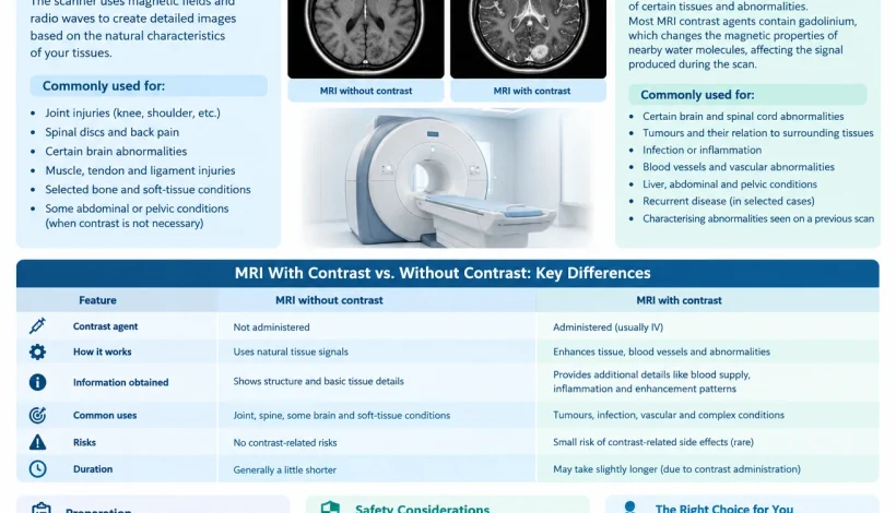
MRI With Contrast vs. Without Contrast

MRI With Contrast vs. Without Contrast: What Is the Difference?

Magnetic Resonance Imaging (MRI) is a medical imaging technique that uses a strong magnetic field and radio waves to produce detailed images of organs, tissues, and other structures inside the body. It is commonly used to investigate neurological conditions, joint injuries, spinal problems, and abnormalities affecting various organs.

When an MRI is recommended, patients may be asked to undergo an examination with contrast or without contrast. The difference lies in whether a contrast agent is administered to improve the visibility of certain tissues, blood vessels, or abnormalities.o

Understanding MRI with contrast vs. without contrast can help patients know what to expect during the examination and why a particular approach may be recommended. However, neither method is universally better. The appropriate examination depends on the medical condition being investigated, the body part being examined, and the information required by the treating doctor.

This article explains how MRI contrast works, when each type of scan may be used, and the preparation and safety considerations patients should understand.

What Is an MRI Scan Without Contrast?

An MRI without contrast is performed without administering an injectable contrast agent. The scanner uses magnetic fields and radio waves to generate images based on the natural characteristics of tissues.

Different tissues produce different signal patterns, allowing radiologists to assess their structure and identify abnormalities. Multiple imaging sequences may be used to provide additional information about the area being examined.

Common Uses of MRI Without Contrast

An MRI without contrast may be recommended for:

  • Assessing certain knee, shoulder, and other joint injuries.
  • Examining spinal discs and investigating selected causes of back pain.
  • Evaluating certain brain abnormalities.
  • Investigating some muscle, tendon, and ligament injuries.
  • Assessing selected bone and soft-tissue conditions.
  • Examining particular abdominal or pelvic structures when contrast is not necessary.

The decision depends on the suspected condition and the examination protocol. Some conditions can be evaluated adequately without contrast, while others require additional information that contrast-enhanced imaging may provide.

Advantages of MRI Without Contrast

MRI without contrast offers several practical advantages:

  • No intravenous contrast injection is required.
  • It avoids risks specifically associated with the administered contrast agent.
  • It can provide detailed images of many soft-tissue structures.
  • It may be sufficient for the diagnostic question being investigated.

An MRI without contrast is not necessarily less informative. Its suitability depends on whether the images can adequately answer the clinical question.

What Is an MRI Scan With Contrast?

An MRI with contrast involves administering a contrast agent, usually through an intravenous line, to improve the visibility of certain tissues and abnormalities.

Most MRI contrast agents contain gadolinium, a metal used in a specially formulated compound. Following administration, the agent changes the magnetic properties of nearby water molecules, affecting the signal produced during MRI.

These changes can help radiologists assess tissue enhancement, blood supply, inflammation, and other characteristics of an abnormality.

Contrast is not required for every MRI examination. It is used when the additional information is expected to be clinically useful.

Common Uses of MRI With Contrast

An MRI with contrast may be recommended for:

  • Evaluating certain brain and spinal cord abnormalities.
  • Assessing selected tumours and their relationship with surrounding tissues.
  • Investigating suspected infection or inflammation.
  • Evaluating certain blood vessels and vascular abnormalities.
  • Assessing some liver, abdominal, and pelvic conditions.
  • Investigating suspected recurrent disease in selected clinical situations.
  • Characterising abnormalities identified on an earlier examination.

For some conditions, doctors may request images both before and after contrast administration. Comparing the images can help identify changes that may not be fully characterised by non-contrast imaging alone.

How Does Contrast Improve MRI Images?

Contrast enhancement occurs when the administered agent changes the signal characteristics of tissues.

Different abnormalities may show different enhancement patterns. For example, certain tumours, inflammatory changes, and areas with altered blood-brain barrier integrity may enhance after contrast administration.

However, enhancement does not automatically indicate cancer or another specific disease. Benign conditions, inflammation, and normal tissue characteristics can also produce enhancement.

Radiologists interpret these patterns alongside other MRI sequences, the patient’s medical history, and relevant clinical findings.

MRI With Contrast vs. Without Contrast: Key Differences

The main difference between these examinations is whether a contrast agent is administered and whether the additional information it provides is needed.

Feature

MRI without contrast

MRI with contrast

Contrast agent

Not administered

Usually administered intravenously

Image information

Shows tissue structure and signal characteristics

Adds information about tissue enhancement and contrast distribution

Common applications

Selected joint, spine, brain, and soft-tissue examinations

Selected tumour, inflammatory, vascular, and post-treatment evaluations

Injection required

Usually not for contrast

Usually yes

Additional safety considerations

MRI magnetic-field safety still applies

MRI safety plus assessment of contrast-related risks

Kidney function assessment

Usually not required solely for a non-contrast examination

May be relevant depending on the contrast agent and the patient’s medical history

Examination duration

Depends on the protocol

May take longer because additional sequences are acquired

Diagnostic suitability

Appropriate when contrast is unnecessary

Appropriate when enhancement information is needed

The exact examination protocol varies according to the body part, suspected condition, scanner, and clinical indication.

When Is MRI With Contrast Recommended?

Contrast-enhanced MRI is considered when information about tissue enhancement may help answer the clinical question.

1. Brain and Neurological Conditions

MRI with contrast may be used to evaluate selected brain tumours, infections, inflammatory conditions, and abnormalities affecting the brain or its coverings.

In some situations, contrast helps distinguish different types of tissue changes or assess the extent of an abnormality.

However, many brain MRI examinations can be performed without contrast, depending on the reason for imaging.

2. Tumour Evaluation

Contrast may help characterise certain tumours by showing how an abnormality enhances and how it relates to surrounding structures.

It can also be useful in selected follow-up examinations when doctors need to evaluate changes after treatment.

Enhancement patterns are not sufficient on their own to establish a diagnosis. The findings may need to be considered alongside other imaging studies, laboratory investigations, or biopsy results.

3. Infection and Inflammation

Certain infections and inflammatory conditions can cause changes in tissue enhancement.

Contrast-enhanced MRI may help assess the location and extent of an abnormality when clinically indicated.

The choice depends on the suspected condition and whether contrast is expected to provide additional diagnostic information.

4. Blood Vessels and Selected Organ Conditions

Some MRI examinations use contrast to assess blood vessels or evaluate abnormalities in organs such as the liver and pelvis.

The appropriate technique depends on the suspected disease and the type of information required. Some vascular MRI examinations can also be performed without contrast using specialised sequences.

When Is MRI Without Contrast Sufficient?

MRI without contrast may provide enough information when the examination is intended to assess structural changes that can be evaluated using standard MRI sequences.

Examples include:

  • Many routine examinations of the knee and shoulder.
  • Assessment of selected spinal disc abnormalities.
  • Certain investigations of back pain.
  • Evaluation of some joint and soft-tissue injuries.
  • Selected brain examinations where contrast is not clinically necessary.

For example, an MRI performed to investigate a suspected ligament injury may not require contrast. However, the decision can change if the doctor is investigating infection, a tumour, or a different underlying condition.

The choice should be based on the clinical question rather than a general preference for one type of MRI.

Is MRI With Contrast Safe?

MRI contrast agents are generally well tolerated when used appropriately, but they are not completely free of risk.

Most gadolinium-based contrast agents have a good safety profile. Nevertheless, patients should inform the imaging team about relevant medical conditions and any previous reaction to an MRI contrast agent.

Possible Side Effects

Some patients may experience mild, temporary effects, including:

  • Nausea.
  • Headache.
  • Dizziness.
  • A feeling of coldness or discomfort during injection.
  • Discomfort at the injection site.

Allergic-like reactions can occur, although severe reactions are uncommon.

A history of a previous reaction to a gadolinium-based contrast agent should be discussed before the examination. The imaging team can assess the circumstances and determine appropriate precautions.

Kidney Function and MRI Contrast

Kidney function is an important consideration for certain patients receiving gadolinium-based contrast agents.

In people with severe kidney impairment, some agents have been associated with a rare condition called nephrogenic systemic fibrosis. The risk depends on the specific agent and the patient’s medical circumstances.

Modern contrast agents differ in their safety profiles. The healthcare team determines whether kidney function assessment is necessary and which agent is appropriate.

Patients should inform the imaging team if they have kidney disease, are receiving dialysis, or have been advised that their kidney function is significantly reduced.

Pregnancy and Breastfeeding

If you are pregnant or may be pregnant, inform your doctor and the imaging team before an MRI examination.

Gadolinium-based contrast is generally avoided during pregnancy unless the expected clinical benefit justifies its use after an individual assessment.

For breastfeeding patients, routine interruption of breastfeeding is generally not necessary after administration of most gadolinium-based contrast agents. However, patients should discuss individual concerns with their healthcare professional.

How to Prepare for an MRI With or Without Contrast

Preparation depends on the body part being examined and the imaging protocol.

Before the Examination

Patients should:

  • Inform the imaging team about pacemakers, implants, and previous surgeries.
  • Report any previous reaction to an MRI contrast agent.
  • Inform staff about kidney disease, pregnancy, or other relevant medical conditions.
  • Follow instructions about eating and drinking.
  • Ask whether any recent blood test or kidney function assessment is required.
  • Remove metal objects and follow instructions regarding clothing and accessories.
  • Share relevant medical records and previous imaging reports when available.

Not every MRI examination requires fasting or blood tests. These requirements depend on the specific examination and the patient’s medical history.

During an MRI Without Contrast

The patient lies on a scanning table that moves into the MRI scanner. The imaging team positions the patient and acquires images using the required sequences.

The scanner makes loud knocking or tapping sounds, so hearing protection is generally provided. Remaining still helps reduce motion-related image artefacts.

During an MRI With Contrast

The examination may begin with images acquired before contrast administration. A contrast agent is then injected through an intravenous line, followed by additional image sequences.

The patient may notice the insertion of the intravenous line or brief discomfort at the injection site. The imaging team monitors the procedure and provides instructions throughout the examination.

The total duration varies according to the body part and the number of sequences required.

Does MRI With Contrast Take Longer?

MRI with contrast may take longer than a comparable examination without contrast because additional images are acquired before and after injection.

However, the difference in duration varies considerably. Some examinations require several specialised sequences, while others are relatively straightforward.

The total appointment time may also include patient preparation, safety screening, positioning, intravenous access, and any necessary post-examination observation.

Patients should follow the appointment instructions provided by the imaging centre.

Frequently Asked Questions

1. Which is better: MRI with contrast or without contrast?

Neither is universally better. MRI without contrast is sufficient for many examinations, while contrast-enhanced MRI may provide additional information for selected tumours, inflammatory conditions, vascular abnormalities, and other clinical indications.

2. Is MRI without contrast less accurate?

Not necessarily. An MRI without contrast can provide highly useful diagnostic information when it is appropriate for the condition being investigated. Accuracy depends on the clinical question, imaging protocol, and characteristics of the abnormality.

3. Can MRI contrast damage the kidneys?

Kidney-related risks depend on the patient’s kidney function and the specific gadolinium-based contrast agent used. Patients with significant kidney impairment should inform the imaging team so that appropriate precautions can be considered.

4. Do all MRI scans require contrast?

No. Many MRI examinations are performed without contrast. The decision depends on the suspected condition and whether contrast is expected to provide useful additional information.

5. Can I eat before an MRI with contrast?

Fasting is not required for every MRI with contrast. Some examinations may have specific food or drink restrictions. Follow the instructions provided for your particular appointment.

6. How long does an MRI with contrast take?

The duration varies by examination. Many MRI scans take approximately 20–60 minutes, although complex examinations may take longer. Contrast administration and additional imaging sequences can affect the total time.

7. Can I have an MRI if I have a pacemaker?

Some pacemakers and implanted devices are compatible with MRI under specific conditions, while others may present risks. Inform the imaging team about the exact device before scheduling the examination. Do not assume that an implant is safe for MRI without appropriate screening.

Conclusion

The difference between MRI with contrast vs. without contrast lies in whether an additional agent is used to improve the visualisation of certain tissues and abnormalities.

MRI without contrast can provide detailed information about many joints, spinal structures, and soft tissues. MRI with contrast may offer additional information when doctors need to assess enhancement patterns associated with selected tumours, infections, inflammatory conditions, or vascular abnormalities.

The appropriate examination depends on the patient’s medical history, the condition being investigated, and the diagnostic information required. Patients should discuss concerns about contrast agents, kidney function, implants, pregnancy, and preparation with their treating doctor and imaging team.

Medical disclaimer: This article is intended for general educational purposes only. It does not replace professional medical advice, diagnosis, or treatment. The decision to perform MRI with or without contrast should be based on individual clinical circumstances and the advice of qualified healthcare professionals.


Name: Ace Imaging Center

Address: Bezolla Complex, 102, VN Purav Marg, Swastik Park, Chembur, Mumbai, Maharashtra 400071

contact: 081693 60334
🌐 https://aceimaging.in

    Book an appointment

    Fill form and get discount. Check Offers


    MRI vs PET Scan vs CT Scan

    MRI vs PET Scan vs CT Scan: Understanding the Key Differences

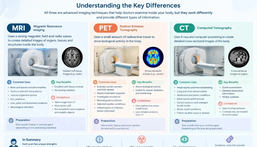
    MRI, PET, and CT scans are medical imaging techniques that help doctors examine internal structures, investigate diseases, and guide treatment decisions. Although all three provide images of the body, they work differently and offer different types of medical information.

    When comparing MRI vs PET scan vs CT scan, it is important to understand what each examination can reveal. MRI provides detailed images of soft tissues, CT produces cross-sectional images of internal structures using X-rays, and PET evaluates metabolic or other biological activity using a radioactive tracer.

    Doctors select an imaging examination according to the patient’s symptoms, medical history, suspected condition, and the information required for diagnosis or treatment planning. In some situations, more than one imaging technique may be recommended because the tests provide complementary information.

    This article explains the differences between MRI, PET, and CT scans, including their uses, preparation requirements, advantages, and limitations.

    What Is an MRI Scan?

    Magnetic Resonance Imaging (MRI) is a medical imaging technique that uses a strong magnetic field and radio waves to create detailed images of organs, tissues, and other structures inside the body.

    Unlike CT scanning, MRI does not use ionising radiation. It is particularly useful for examining soft tissues, including the brain, spinal cord, muscles, ligaments, and certain internal organs.

    Common Uses of MRI

    Doctors may recommend an MRI scan to investigate:

    • Brain and spinal cord abnormalities.
    • Certain tumours and soft-tissue lesions.
    • Ligament, tendon, and joint injuries.
    • Intervertebral disc problems.
    • Selected liver, pelvic, and abdominal conditions.
    • Certain neurological disorders.
    • Changes in soft tissues that require detailed assessment.

    The specific type of MRI depends on the area being examined and the clinical question. Some examinations use a contrast agent to improve the visibility of particular tissues or abnormalities.

    Advantages and Limitations of MRI

    MRI provides excellent soft-tissue contrast and does not involve ionising radiation. However, the examination usually takes longer than a CT scan, and patients must remain relatively still during imaging.

    The strong magnetic field also means that certain implants and metallic objects require careful assessment before the examination. Patients should inform the imaging team about pacemakers, implants, previous surgeries, and any possibility of metallic fragments in the body.

    What Is a PET Scan?

    Positron Emission Tomography (PET) is a type of nuclear medicine imaging that evaluates biological activity within the body. It uses a radiotracer, which is a small amount of radioactive material administered according to the examination protocol.

    One commonly used tracer is fluorodeoxyglucose (FDG), a glucose-like substance that helps demonstrate glucose metabolism in tissues.

    After administration, the tracer travels through the body and accumulates according to the biological processes being assessed. The scanner detects signals from the tracer and generates images that show its distribution.

    Common Uses of PET Scans

    PET imaging may be used to:

    • Evaluate certain cancers and their spread.
    • Assess treatment response in selected cancers.
    • Investigate suspected cancer recurrence.
    • Examine certain neurological conditions.
    • Evaluate selected cardiac conditions.
    • Investigate specific inflammatory or infectious conditions when indicated.

    PET findings show patterns of biological activity, not cancer alone. Infection, inflammation, and normal physiological processes can also cause increased tracer uptake.

    What Is a PET-CT Scan?

    A PET-CT scan combines PET imaging with Computed Tomography. The PET component provides information about metabolic activity, while the CT component shows the anatomical location of that activity.

    This combination can help doctors assess whether an area of increased tracer uptake corresponds to a particular organ, lymph node, or other structure.

    PET-CT is commonly used in oncology, although its suitability depends on the suspected disease and the clinical purpose of the examination.

    What Is a CT Scan?

    Computed Tomography (CT) uses X-rays and computer processing to create detailed cross-sectional images of the body.

    Unlike a conventional X-ray, which produces a projection image, CT provides multiple images that can be reconstructed into views of internal structures from different angles.

    CT is frequently used when doctors need to examine organs, bones, blood vessels, or internal injuries. It can also be used to investigate certain cancers and support treatment planning.

    Common Uses of CT Scans

    A CT scan may be recommended to investigate:

    • Head injuries and suspected internal bleeding.
    • Lung and chest abnormalities.
    • Abdominal and pelvic conditions.
    • Bone injuries and complex fractures.
    • Certain tumours and enlarged lymph nodes.
    • Selected blood vessel conditions.
    • Changes in organs following injury or disease.

    Some CT examinations use an iodine-based contrast agent to improve the visibility of blood vessels, organs, or other structures. Contrast is not required for every CT scan.

    Advantages and Limitations of CT

    CT examinations are generally quick and provide detailed anatomical information. This makes CT particularly useful in emergency settings and when rapid assessment is necessary.

    However, CT uses ionising radiation. Contrast-enhanced examinations also require consideration of previous contrast reactions, kidney function when relevant, and other patient-specific factors.

    The potential benefits and risks should be considered when selecting the appropriate examination.

    MRI vs PET Scan vs CT Scan: Key Differences

    The main difference between these imaging techniques is the type of information they provide.

    Feature MRI scan PET scan CT scan
    Full form Magnetic Resonance Imaging Positron Emission Tomography Computed Tomography
    Imaging method Magnetic field and radio waves Radioactive tracer and specialised detector X-rays and computer processing
    Primary information Detailed anatomy and soft-tissue characteristics Metabolic or other biological activity Detailed cross-sectional anatomy
    Ionising radiation No Yes, from the radiotracer Yes
    Common applications Brain, spine, joints, soft tissues, selected organs Selected cancers, treatment assessment, neurological and cardiac applications Trauma, chest, abdomen, bones, and selected cancers
    Contrast or tracer Contrast may be used Radiotracer is generally required Contrast may be used
    Typical examination time Often around 20–60 minutes, depending on protocol Usually longer because tracer uptake takes time Often completed within minutes
    Important consideration Metal implants and magnetic-field safety Tracer-specific preparation and interpretation Radiation exposure and possible contrast-related risks

    These are general comparisons. Actual examination times, preparation requirements, and diagnostic usefulness vary according to the body part, protocol, and clinical indication.

    MRI vs PET Scan vs CT Scan for Cancer

    MRI, PET, and CT scans can all contribute to cancer assessment, but they answer different clinical questions.

    MRI for Cancer Evaluation

    MRI can provide detailed information about the location and extent of abnormalities in soft tissues. Depending on the type of cancer, it may help assess involvement of nearby structures and support treatment planning.

    For example, MRI is frequently used in the evaluation of selected brain, spinal, pelvic, and musculoskeletal tumours.

    PET-CT for Cancer Evaluation

    PET-CT can provide information about metabolic activity and its anatomical location. In selected cancers, it may help assess disease distribution, treatment response, or suspected recurrence.

    However, not every cancer demonstrates high uptake of a particular PET tracer. Small lesions and some tumours with low tracer uptake may be difficult to detect.

    CT for Cancer Evaluation

    CT can show the size, location, and anatomical relationships of tumours and may help assess lymph nodes, lungs, abdominal organs, and other structures.

    It is commonly used in cancer diagnosis, staging, and follow-up when clinically appropriate.

    Important: No single imaging test is suitable for every type of cancer. The choice depends on the suspected cancer, the clinical question, and the information needed by the treating team.

    MRI vs PET Scan vs CT Scan for Brain Conditions

    The preferred examination for a brain condition depends on the symptoms and the suspected cause.

    MRI: MRI provides detailed images of brain tissue and is frequently used to investigate certain tumours, neurological disorders, inflammation, and abnormalities involving the brain or spinal cord.

    CT: CT can rapidly identify certain urgent problems, including intracranial bleeding and skull fractures. It may be selected when immediate imaging is required.

    PET: Certain PET examinations can assess brain metabolism or other biological processes. They may be used in selected neurological evaluations, depending on the tracer and clinical indication.

    These tests are not interchangeable. For sudden neurological symptoms, the treating team determines which examination is appropriate based on the urgency and suspected condition.

    Which Scan Is More Accurate: MRI, PET, or CT?

    There is no universal answer to which scan is more accurate. Diagnostic performance depends on the disease being investigated, the body part examined, the imaging protocol, and the characteristics of the abnormality.

    MRI may provide more useful detail for certain soft-tissue conditions, while CT may be more appropriate for rapid assessment of particular injuries. PET-CT may provide additional information when metabolic activity is important to the clinical question.

    A scan can also produce inconclusive, false-positive, or false-negative findings. For example, inflammation may cause increased uptake on PET imaging, while some small lesions may not be clearly visible.

    Doctors interpret imaging results alongside symptoms, physical examination findings, laboratory tests, previous images, and other investigations. Additional testing may be necessary when the findings do not adequately explain the clinical picture.

    Radiation Exposure: MRI vs PET Scan vs CT Scan

    Radiation exposure is another important difference between these imaging techniques.

    MRI Radiation Exposure

    MRI does not use ionising radiation. However, it involves a strong magnetic field, and the safety of implants and other metallic objects must be assessed before scanning.

    PET Scan Radiation Exposure

    PET imaging uses a radioactive tracer. The amount of radiation depends on the tracer, the administered activity, and the examination protocol.

    When PET is combined with CT, the CT component also contributes to the total radiation exposure.

    CT Scan Radiation Exposure

    CT uses X-rays to produce cross-sectional images. Radiation exposure varies according to the body part, scan settings, and examination protocol.

    Doctors consider the expected diagnostic benefit when recommending imaging that involves ionising radiation. Patients who are pregnant or may be pregnant should inform their healthcare team before the examination.

    How to Prepare for MRI, PET, and CT Scans

    Preparation requirements vary according to the examination and the patient’s medical circumstances.

    Scan type General preparation considerations
    MRI Inform staff about implants, pacemakers, metallic objects, previous surgeries, and claustrophobia. Follow instructions regarding contrast and clothing.
    PET or PET-CT Follow tracer-specific fasting instructions, discuss diabetes and medications, and follow guidance regarding physical activity and hydration.
    CT Follow instructions regarding food, fluids, and contrast when applicable. Report previous contrast reactions, relevant medical conditions, and possible pregnancy.

    Patients should not independently stop medication or change prescribed doses before an examination.

    For all three imaging techniques, it is helpful to bring relevant medical records, previous imaging reports, and the referral information provided by the treating doctor.

    Frequently Asked Questions

    1. What is the main difference between MRI, PET, and CT scans?

    MRI uses magnetic fields and radio waves to produce detailed images, CT uses X-rays to show internal anatomy, and PET uses a radioactive tracer to evaluate biological activity. PET-CT combines metabolic and anatomical information.

    2. Is MRI safer than a CT scan?

    MRI does not use ionising radiation, whereas CT does. However, safety depends on individual circumstances, including the need for diagnostic information, MRI implant compatibility, and any contrast-related considerations. The appropriate examination should be selected according to clinical need.

    3. Is a PET scan the same as a CT scan?

    No. PET assesses biological activity using a radiotracer, while CT provides anatomical images using X-rays. A PET-CT examination combines the two techniques.

    4. Can MRI, PET, and CT scans detect cancer?

    All three techniques can contribute to cancer assessment in appropriate situations. Their usefulness depends on the cancer type, location, size, and clinical question. Imaging findings alone may not establish a diagnosis, and biopsy or other investigations may be required.

    5. Which scan takes the least time?

    CT scanning itself is often completed within minutes. MRI commonly takes longer, while PET examinations require additional time for tracer administration and uptake before image acquisition. Actual duration varies by protocol.

    6. Can a patient need more than one type of scan?

    Yes. Different imaging techniques may provide complementary information. A doctor may recommend more than one examination when the findings from a single test are insufficient to answer the clinical question.

    7. Does every PET scan include a CT scan?

    No. PET can be performed as a standalone examination, although PET-CT is widely used. The appropriate technique depends on the clinical indication and available protocol.

    Conclusion

    MRI, PET, and CT scans serve different purposes in medical imaging. MRI provides detailed soft-tissue images without ionising radiation, CT offers rapid and detailed anatomical assessment using X-rays, and PET evaluates biological activity using a radioactive tracer.

    Understanding the differences between MRI vs PET scan vs CT scan can help patients understand why different examinations may be recommended. However, the choice should be based on the patient’s symptoms, medical history, suspected condition, and the diagnostic information required.

    Patients should discuss questions about preparation, contrast agents, radiation exposure, and examination suitability with their treating doctor and imaging team.

    Medical disclaimer: This article is intended for general educational purposes and does not replace professional medical advice, diagnosis, or treatment. The selection and interpretation of medical imaging examinations should be based on individual clinical circumstances and the advice of qualified healthcare professionals.


    Name: Ace Imaging Center

    Address: Bezolla Complex, 102, VN Purav Marg, Swastik Park, Chembur, Mumbai, Maharashtra 400071

    contact: 081693 60334
    🌐 https://aceimaging.in

      Book an appointment

      Fill form and get discount. Check Offers


      What Can Affect PET Scan Accuracy

      What Can Affect PET Scan Accuracy? Factors Patients Should Know

      CT Scan Price in India

      A Positron Emission Tomography (PET) scan is a medical imaging examination used to evaluate metabolic activity within the body. It can help doctors investigate certain cancers, assess treatment response, and evaluate selected neurological, cardiac, and inflammatory conditions. When combined with Computed Tomography (CT), a PET-CT scan provides functional and anatomical information that supports clinical assessment.

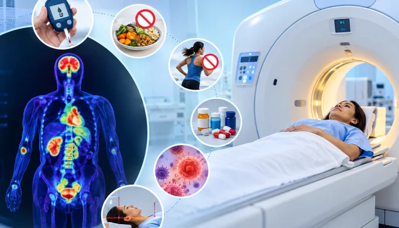
      However, PET scan findings can be influenced by several factors, including blood glucose levels, food intake, physical activity, medications, inflammation, and the characteristics of the condition being investigated. Understanding these factors helps patients prepare appropriately and understand the limitations of the examination.

      What can affect PET scan accuracy? The answer depends on the tracer used, the patient’s medical condition, the purpose of the examination, and the technical quality of the images. Although some factors can be managed through preparation, others are related to the biological characteristics of the disease or the limitations of medical imaging.

      What Is a PET Scan and How Does It Work?

      A PET scan uses a radiotracer, which is a small amount of a radioactive substance, to examine biological processes within the body. One commonly used tracer is fluorodeoxyglucose (FDG), a glucose-like substance that helps demonstrate how tissues use glucose.

      After the tracer is injected into a vein, it travels through the bloodstream and accumulates in tissues according to their metabolic activity. The scanner detects signals from the tracer and creates images showing its distribution.

      A PET-CT examination combines these functional images with CT images, helping doctors identify the anatomical location of areas showing altered metabolic activity.

      PET scans may be used to:

      • Investigate and stage certain cancers.
      • Assess whether cancer has spread to other parts of the body.
      • Evaluate metabolic changes during or after treatment.
      • Investigate suspected recurrence of certain cancers.
      • Assess selected neurological and cardiac conditions.
      • Evaluate specific infections or inflammatory conditions when indicated.

      Increased tracer uptake does not automatically indicate cancer, and a scan without an obvious abnormality does not exclude every disease. Results must be interpreted alongside the patient’s symptoms, medical history, physical examination, and other investigations.

      What Can Affect PET Scan Accuracy?

      Several patient-related, biological, and technical factors can influence PET scan findings. The importance of each factor varies according to the tracer and imaging protocol used.

      1. Blood Glucose Levels

      Blood glucose levels can influence the distribution of FDG during a PET scan. Because FDG behaves similarly to glucose, elevated blood glucose can compete with the tracer and alter its uptake in different tissues.

      High glucose levels may affect image interpretation and the visibility of certain abnormalities. The degree of interference depends on the blood glucose level, the clinical indication, and the imaging protocol.

      Patients with diabetes may require specific preparation instructions because blood glucose management and the timing of insulin administration can influence FDG distribution.

      To prepare appropriately:

      • Follow the fasting instructions provided by the imaging centre.
      • Inform the medical team if you have diabetes.
      • Share information about your diabetes medicines and usual glucose levels.
      • Follow specific instructions about insulin and other glucose-lowering medications.
      • Inform the staff if your blood glucose is unusually high or low.

      Medication doses should not be changed independently. The treating doctor and imaging team can determine whether any adjustments or changes in scan timing are necessary.

      2. Eating or Drinking Before the Examination

      Food intake before an FDG PET scan may affect insulin levels and glucose metabolism. Eating shortly before the examination, particularly carbohydrate-rich foods, can alter tracer uptake in muscles and other tissues.

      For many FDG PET scans, patients are instructed to fast for approximately four to six hours. However, fasting requirements depend on the tracer, examination protocol, and individual medical circumstances.

      Plain water is generally permitted and often encouraged, unless different instructions have been provided.

      Patients should confirm whether beverages such as tea, coffee, milk, juice, and other caloric drinks are permitted before their appointment.

      Following the prescribed preparation instructions helps reduce avoidable changes in tracer distribution.

      3. Recent Physical Activity

      Physical activity increases glucose utilisation in working muscles. Since FDG uptake is influenced by glucose metabolism, strenuous exercise before a scan can lead to increased tracer accumulation in muscles.

      This may make some areas more difficult to interpret or affect the appearance of surrounding tissues.

      Patients are commonly advised to avoid strenuous exercise for approximately 24 hours before an FDG PET scan, or for the period specified by the imaging team.

      Preparation may include:

      • Avoiding vigorous workouts and heavy physical exertion.
      • Following instructions about running, cycling, and other strenuous activities.
      • Resting quietly during the tracer uptake period.
      • Informing the staff about recent intense physical activity.

      The recommended restrictions may vary according to the examination and the patient’s medical circumstances.

      4. Medications and Recent Medical Treatments

      Certain medications can influence tracer distribution or the appearance of tissues on PET images.

      For example, insulin administered too close to an FDG PET scan may increase tracer uptake in muscles. Other medicines that affect glucose metabolism or inflammation may also influence findings, depending on the clinical situation.

      Recent chemotherapy, radiotherapy, immunotherapy, surgery, or other medical procedures can cause changes in tissue metabolism. These changes may complicate the interpretation of findings when treatment effects resemble disease activity.

      Patients should inform the imaging team about:

      • Prescription and non-prescription medicines.
      • Insulin and other diabetes treatments.
      • Recent chemotherapy or radiotherapy.
      • Immunotherapy and other cancer-related treatments.
      • Recent surgery, biopsies, or invasive procedures.
      • Recent changes in medication.

      Patients should not stop prescribed medication without medical advice. The timing of the examination should be determined according to the clinical question and the relevant imaging protocol.

      5. Infection and Inflammation

      PET scans identify metabolic activity rather than cancer alone. Activated inflammatory cells can also accumulate FDG, producing areas of increased tracer uptake.

      Infections, inflammatory disorders, and healing tissues may therefore produce findings that resemble malignancy.

      For example, an inflamed lymph node may demonstrate increased uptake even when the cause is not cancer. Similarly, inflammation around a recent surgical site may affect the appearance of the region being examined.

      The radiologist considers the pattern and location of uptake alongside the patient’s symptoms, medical history, laboratory results, and other imaging findings.

      Patients should inform their doctor about recent infections, fever, inflammatory conditions, injuries, or surgical procedures before the examination.

      6. Movement During Image Acquisition

      Movement during a PET-CT scan can reduce image quality. It may cause blurring, misalignment between PET and CT images, or difficulty identifying the precise location of an abnormality.

      Movement may occur because of discomfort, coughing, anxiety, involuntary muscle activity, or difficulty remaining in one position.

      Patients can help minimise movement by following positioning instructions, wearing suitable clothing, and informing the staff about pain or physical limitations.

      If movement significantly affects image quality, additional imaging may sometimes be necessary.

      7. Timing Between Tracer Injection and Scanning

      The interval between tracer administration and image acquisition is an important part of PET scan preparation.

      After injection, the tracer requires time to distribute through the body. For many FDG PET scans, image acquisition begins approximately 60 minutes after injection, although the precise interval varies by protocol.

      The timing affects tracer distribution and the appearance of tissues. Imaging outside the intended protocol may make comparisons with previous examinations more difficult.

      The imaging team determines the appropriate uptake period based on the tracer, the clinical indication, and the examination protocol.

      8. Size and Location of an Abnormality

      The size and location of a lesion can influence whether it is visible on a PET scan.

      Very small abnormalities may be difficult to detect because of the scanner’s spatial resolution. The partial-volume effect can also cause tracer uptake in small lesions to appear lower than it actually is.

      Some tumours demonstrate relatively low FDG uptake, which may make them less conspicuous. Other lesions may be difficult to distinguish because of normal tracer activity in nearby tissues.

      For example, normal physiological uptake in the brain, heart, kidneys, and urinary tract can affect the visibility of abnormalities in or around these structures.

      A negative PET scan does not exclude every type of cancer or disease. Depending on the clinical circumstances, further imaging, laboratory testing, or biopsy may be necessary.

      9. Body Characteristics and Image Quality

      Body size and tissue characteristics can influence image noise and the visibility of smaller abnormalities.

      In some patients, greater tissue thickness can reduce image clarity. Modern scanners and image reconstruction techniques help address these limitations, but they cannot eliminate every source of image noise.

      Body composition and the distribution of muscle and fat may also influence tracer uptake.

      These factors do not automatically mean that a scan is inaccurate. Instead, they are considered when assessing the quality of the images and interpreting the findings.

      10. Scanner Performance and Imaging Protocol

      PET scan interpretation depends partly on the technical quality of the images.

      Factors that may influence image quality include:

      • Scanner calibration and maintenance.
      • Appropriate tracer administration.
      • Image acquisition duration.
      • Patient positioning.
      • Attenuation correction and image reconstruction.
      • Standardised imaging protocols.
      • The availability of relevant previous scans.

      Quality assurance procedures and appropriate technical protocols help reduce avoidable imaging errors. However, technical quality alone cannot overcome every biological limitation of a PET scan.

      11. Medical History and Previous Imaging

      Clinical information is important when interpreting PET scan findings.

      Details about previous diagnoses, symptoms, treatments, surgery, infections, and other investigations can help the radiologist assess the significance of an abnormal area.

      Previous imaging studies may also help determine whether a finding is new, unchanged, or has changed over time.

      Patients should provide relevant medical records and inform the imaging team about their clinical history. The referring doctor and radiologist can use this information to interpret the results in context.

      Factors That Can Affect PET Scan Accuracy: Summary

      Factor

      Potential influence on findings

      Elevated blood glucose

      May alter FDG distribution and uptake

      Food intake

      May affect insulin levels and glucose metabolism

      Strenuous exercise

      May increase tracer uptake in muscles

      Medications

      May influence tracer distribution or tissue metabolism

      Infection or inflammation

      May cause increased uptake unrelated to cancer

      Recent surgery or treatment

      May produce healing-related or treatment-related changes

      Movement

      May cause image blurring or misalignment

      Small lesion size

      May reduce visibility because of spatial resolution

      Scanner performance

      May affect image clarity and detection

      Incomplete medical history

      May make findings more difficult to interpret in context

      How Should Patients Prepare for a PET Scan?

      Preparation requirements depend on the tracer and examination being performed. Patients should follow the instructions provided by the imaging team rather than relying on general advice alone.

      Common preparation considerations include:

      • Fasting: Confirm when to stop eating and which drinks are permitted.
      • Diabetes management: Obtain specific instructions regarding blood glucose monitoring and medication timing.
      • Physical activity: Follow the recommended restrictions on strenuous exercise.
      • Medication history: Share information about prescribed medicines and recent treatment.
      • Medical history: Report recent infections, surgery, biopsies, or other relevant procedures.
      • Hydration: Drink water as instructed.
      • Clothing: Follow guidance regarding comfortable clothing and metal accessories.
      • Previous reports: Bring relevant medical records and earlier imaging studies, when available.
      • Pregnancy or breastfeeding: Inform the medical team beforehand, if applicable, so the examination can be assessed appropriately.

      If any preparation instruction is unclear, patients should contact the imaging team before the examination.

      Can a PET Scan Produce False-Positive or False-Negative Findings?

      Yes. PET scans have diagnostic limitations and can produce false-positive or false-negative findings.

      A false-positive finding occurs when an area appears suspicious even though the suspected disease is not present. Inflammation, infection, and healing after surgery may contribute to this type of finding.

      A false-negative finding occurs when a disease or abnormality is present but is not detected by the scan. Small lesion size, low tracer uptake, and other imaging limitations may contribute.

      The significance of a finding depends on the clinical context. Doctors may recommend additional imaging, laboratory tests, follow-up examinations, or biopsy when necessary.

      PET scan results should not be used in isolation to establish or exclude every diagnosis. They are interpreted alongside other relevant clinical information.

      When May a Repeat PET Scan Be Necessary?

      A repeat PET scan may be considered when the original images are technically inadequate, preparation requirements were not met, or the examination did not sufficiently answer the clinical question.

      Repeat imaging may also be used when clinically indicated to assess treatment response or investigate suspected disease recurrence.

      However, an unexpected result does not automatically mean that another scan is required. The doctor may first review the original images, compare previous studies, or recommend a different investigation.

      PET and PET-CT examinations involve ionising radiation from the radiotracer and, where applicable, the CT component. Repeat imaging should therefore be based on clinical need and an appropriate assessment of potential benefits and risks.

      Frequently Asked Questions

      1. What can affect PET scan accuracy the most?

      For FDG PET scans, blood glucose levels, fasting, physical activity, medication timing, and the interval between tracer injection and image acquisition are important factors. Inflammation, lesion characteristics, and image quality can also influence interpretation.

      2. Can high blood sugar affect PET scan results?

      Yes. Elevated blood glucose can alter FDG uptake and distribution. Patients with diabetes should follow the specific preparation and medication instructions provided by their healthcare team.

      3. Can eating before a PET scan affect the results?

      Yes. Eating before an FDG PET scan may affect insulin levels and tracer distribution. Many protocols require fasting for several hours, but the exact instructions depend on the examination.

      4. Can inflammation look like cancer on a PET scan?

      Inflammation and certain infections can cause increased tracer uptake that resembles cancer. Additional clinical information or investigations may be required to determine the cause.

      5. Can a PET scan miss cancer?

      Yes. Some small lesions or tumours with low FDG uptake may not be clearly visible. A negative PET scan does not exclude every type of cancer, particularly when clinical suspicion remains.

      6. Should I stop my medication before a PET scan?

      Do not stop or change prescribed medication without medical guidance. Inform the imaging team about all medicines so that appropriate preparation instructions can be provided.

      7. Is a PET-CT scan always accurate?

      No diagnostic imaging examination is completely accurate. PET-CT provides functional and anatomical information, but its findings must be interpreted alongside the patient’s medical history and other relevant investigations.

      Conclusion

      PET scan accuracy depends on several factors, including blood glucose levels, fasting, physical activity, medications, inflammation, recent treatment, movement, lesion characteristics, and technical image quality.

      Following the prescribed preparation instructions and providing accurate medical information can help reduce avoidable factors that affect image interpretation. Understanding the limitations of PET imaging is equally important, as neither increased nor decreased tracer uptake establishes a diagnosis on its own.

      The findings should be interpreted by qualified medical professionals in the context of the patient’s symptoms, medical history, and other investigations.

      Medical disclaimer: This article is intended for general educational purposes only. It does not replace individual medical advice, diagnosis, or treatment. PET scan preparation and interpretation may vary according to the tracer used, the clinical indication, and the patient’s medical condition. Patients should follow the instructions provided by their treating doctor and imaging team.


      Name: Ace Imaging Center

      Address: Bezolla Complex, 102, VN Purav Marg, Swastik Park, Chembur, Mumbai, Maharashtra 400071

      contact: 081693 60334

      🌐 https://aceimaging.in

        Book an appointment

        Fill form and get discount. Check Offers


        MRI Price in India

        MRI Price in India: Complete Guide to MRI Cost, Types and What to Expect

        CT Scan Price in India

        If your doctor has recommended an MRI, one of the first questions you may have is, “What is the MRI price in India?” The answer depends on several factors, including the body part being scanned, the type of MRI examination, whether contrast is required, the number of areas being examined, the imaging protocol and the diagnostic centre.

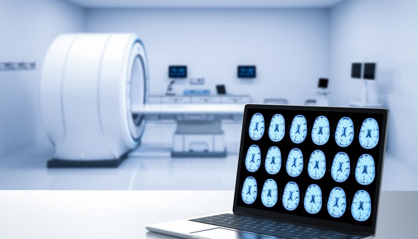
        Magnetic Resonance Imaging (MRI) is an advanced diagnostic imaging technique that uses a powerful magnetic field, radio waves and computer processing to create detailed images of structures inside the body. Unlike X-rays and CT scans, MRI does not use ionising radiation.

        MRI is commonly used to examine the brain, spine, joints, muscles, soft tissues, abdomen, pelvis and blood vessels. Since different MRI examinations require different protocols, there is no single fixed price applicable to every patient.

        Understanding the factors that affect the MRI price in India can help patients compare costs, prepare for the examination and ask the right questions before booking an appointment.

        What Is the MRI Price in India?

        The MRI price in India can vary widely depending on the examination and location.

        One current healthcare cost guide gives an indicative national average of approximately ₹6,000, with an indicative range of around ₹5,100 in some Tier 2 cities and ₹7,500 in Tier 1 cities. These are broad estimates rather than fixed rates.

        Actual prices can differ considerably between individual examinations. For example, current Mumbai listings show MRI prices starting around ₹2,900 for some examinations, while specialised examinations can cost considerably more.

        Some currently listed Mumbai examples include:

        MRI Examination

        Example Price

        MRI Knee – Plain

        Around ₹3,750

        MRI Lumbar Spine – Plain

        Around ₹3,650

        MRI Abdomen & Pelvis

        Around ₹5,500

        MRI Brain with MRA & MRV

        Around ₹6,650

        MRI Cardiac

        Around ₹9,500

        MRI Abdomen Angiogram – Non-Contrast

        Around ₹9,000

        These are examples from current published Mumbai listings and should not be treated as fixed prices for all diagnostic centres.

        A separate Mumbai diagnostic listing shows brain MRI prices of approximately ₹4,000 for a plain examination and ₹6,500 for a contrast examination, further demonstrating how pricing can vary by centre and protocol.

        For the most accurate price, patients should provide the imaging centre with the exact MRI prescription.

        MRI Price in India by Type of Scan

        The type of MRI prescribed is one of the most important factors affecting the total cost.

        Common MRI examinations include:

        Type of MRI

        Commonly Used For

        Cost Consideration

        MRI Brain

        Brain and neurological evaluation

        Depends on protocol and contrast

        MRI Spine

        Spine, discs and surrounding structures

        Varies by spinal region

        MRI Knee

        Ligaments, cartilage, tendons and joint structures

        Plain and contrast prices can differ

        MRI Shoulder

        Shoulder joint and soft tissues

        Depends on protocol

        MRI Abdomen

        Abdominal organs and tissues

        May require specialised sequences

        MRI Pelvis

        Pelvic organs and surrounding structures

        Cost varies by protocol

        MRI Whole Body

        Imaging multiple body regions

        Generally more expensive than single-region MRI

        MRI Angiography

        Blood vessels

        May be contrast or non-contrast

        Cardiac MRI

        Heart and related structures

        Specialised examination

        MRI with Contrast

        Selected conditions requiring enhanced imaging

        Usually costs more than plain MRI

        The exact examination should be based on your doctor’s recommendation and the clinical question that needs to be answered.

        What Factors Affect MRI Price in India?

        1. Body Part Being Examined

        An MRI of a single knee is different from an MRI of the abdomen, brain or multiple spinal regions.

        For example, a current Mumbai listing shows MRI knee and lumbar spine examinations at around ₹3,650–₹3,750, while more specialised examinations such as cardiac MRI are listed at around ₹9,500.

        This is why asking for the price of an unspecified “MRI scan” may not provide an accurate estimate.

        2. MRI With or Without Contrast

        MRI examinations can be performed with or without a contrast agent depending on the medical indication.

        A contrast-enhanced MRI may help make certain tissues, blood vessels or abnormalities more clearly visible. Because contrast involves additional medication and administration, it can increase the overall cost.

        For example, one current Mumbai listing shows a right-knee MRI with contrast starting at approximately ₹6,394, while an examination of both knees with contrast starts at approximately ₹8,075.

        However, contrast is not required for every MRI.

        3. Number of Body Regions

        The price can also change depending on whether one or multiple regions are scanned.

        Examples include:

        • MRI cervical spine
        • MRI lumbar spine
        • MRI cervical and lumbar spine
        • MRI whole spine
        • MRI abdomen
        • MRI abdomen and pelvis
        • MRI multiple joints

        A more extensive examination generally involves additional imaging and reporting.

        4. Specialised MRI Protocols

        Some MRI examinations require specialised protocols depending on the suspected condition.

        Examples include:

        • MR Angiography
        • MR Venography
        • Cardiac MRI
        • Diffusion-weighted MRI
        • Functional MRI
        • Contrast-enhanced MRI
        • Specialised musculoskeletal MRI

        These examinations can have different pricing from routine MRI scans.

        5. Location of the Diagnostic Centre

        MRI prices can vary between cities and even between diagnostic centres within the same city.

        Infrastructure, equipment, staffing, radiologist reporting, operational costs and the type of MRI services offered can all contribute to price differences.

        A national cost estimate currently lists an indicative MRI price of around ₹7,500 for several Tier 1 cities and around ₹5,100 for selected Tier 2 cities, although actual prices can differ significantly.

        MRI Price in India: Plain vs Contrast MRI

        One of the most common questions patients ask is whether they need an MRI with contrast.

        What is a plain MRI?

        A plain MRI is performed without an injected contrast agent. Many routine MRI examinations can provide detailed diagnostic information without contrast.

        It may be used for several examinations involving:

        • Brain
        • Spine
        • Knee
        • Shoulder
        • Ankle
        • Other joints
        • Selected abdominal and pelvic conditions

        What is an MRI with contrast?

        Contrast-enhanced MRI involves administering a contrast agent, generally through an intravenous line, when additional information is required.

        Contrast may be considered for selected cases involving:

        • Tumours or suspected abnormal growths
        • Inflammation
        • Certain infections
        • Blood vessels
        • Post-treatment assessment
        • Specific neurological conditions
        • Selected abdominal or pelvic conditions

        The decision to use contrast should be based on the patient’s clinical requirements.

        Why Does MRI With Contrast Cost More?

        A contrast MRI can have a higher price because it may involve:

        • Contrast material
        • Intravenous administration
        • Additional imaging sequences
        • Additional staff and monitoring
        • More extensive image interpretation

        However, the difference in price varies between examinations and diagnostic centres.

        Patients should ask whether the quoted MRI price includes contrast if contrast has been prescribed.

        What Is Included in the MRI Price?

        Before booking an MRI, ask the diagnostic centre what is included in the quoted price.

        Depending on the centre, the price may include:

        • MRI scanning
        • Technologist assistance
        • Image acquisition
        • Image processing
        • Radiologist reporting
        • Digital images
        • Contrast administration, where applicable

        Additional charges may apply for certain specialised examinations or services.

        Questions to ask before booking

        • Is this price for the exact MRI prescribed by my doctor?
        • Is it a plain or contrast MRI?
        • Is contrast included?
        • Is the radiologist’s report included?
        • Are there any additional charges?
        • When will the report be available?
        • Will I receive digital images?
        • Do I need to fast?
        • Do I need any blood tests before contrast?

        These questions can help you understand the actual cost rather than comparing advertised prices that may cover different examinations.

        How Long Does an MRI Scan Take?

        The duration of an MRI depends on the body part and examination protocol.

        For example, one current listing for an MRI chest states that the examination can take approximately 40–60 minutes.

        A patient’s total appointment may take longer because it can include:

        1. Registration
        2. MRI safety screening
        3. Changing clothes, if required
        4. Removing metallic objects
        5. Positioning
        6. Contrast preparation, if required
        7. Scanning
        8. Post-scan procedures, if applicable

        Patients should therefore allow enough time for the complete appointment.

        How to Prepare for an MRI Scan

        Preparation depends on the type of MRI being performed.

        For many routine MRI examinations, extensive preparation may not be necessary. However, certain examinations may require fasting or other specific instructions.

        Before your MRI:

        • Follow the preparation instructions provided by the imaging centre.
        • Inform the staff about implants or medical devices.
        • Tell the MRI team about previous surgeries.
        • Inform the centre if you have metallic fragments in your body.
        • Tell the staff if you have kidney problems.
        • Inform them about any previous reaction to contrast.
        • Tell the imaging team if you are pregnant or may be pregnant.
        • Remove jewellery, watches and other metallic objects.
        • Bring your doctor’s prescription and previous reports if requested.

        Always follow the specific preparation instructions given for your examination.

        Is MRI Safe?

        MRI does not use ionising radiation. Instead, it uses a strong magnetic field and radio waves to produce images.

        However, MRI safety screening is essential because the magnetic field can interact with certain metallic objects, implants and medical devices.

        Patients should tell the MRI team about:

        • Pacemakers or implanted cardiac devices
        • Metal implants
        • Cochlear implants
        • Surgical clips
        • Neurostimulators
        • Metallic fragments
        • Previous surgeries
        • Other implanted medical devices

        The MRI team can determine whether the examination can be performed safely and under what conditions.

        Is MRI With Contrast Safe?

        MRI contrast is used only when it is considered appropriate for the examination.

        Patients should tell their doctor or imaging team if they have kidney disease or have previously experienced a reaction to MRI contrast.

        The radiology team can assess the relevant medical history and determine whether contrast is appropriate.

        MRI vs CT Scan: What Is the Difference?

        MRI and CT are different imaging techniques and are used for different clinical purposes.

        Feature

        MRI

        CT

        Main technology

        Magnetic field and radio waves

        X-rays

        Ionising radiation

        No

        Yes

        Particularly useful for

        Soft tissues, brain, spine, joints

        Bones, lungs, trauma and many emergency conditions

        Typical scan environment

        Tunnel-like scanner

        Shorter, wider scanner

        Contrast

        Used in selected examinations

        Used in selected examinations

        Scan duration

        Often longer

        Often faster

        The choice between MRI and CT should be made according to the clinical situation and the information your doctor needs.

        How to Choose an MRI Centre

        When considering the MRI price in India, cost is an important factor, but patients should also consider the overall diagnostic service.

        Before booking, check:

        • Whether the centre performs the required MRI
        • Availability of the appropriate MRI protocol
        • Patient safety procedures
        • Equipment and technology
        • Radiologist reporting
        • Image quality
        • Report turnaround time
        • Transparency of pricing
        • Whether previous scans can be provided for comparison

        A lower advertised price may not represent the same examination or services as a higher quoted price, so patients should compare like-for-like MRI examinations.

        Frequently Asked Questions About MRI Price in India

        What is the average MRI price in India?

        A current indicative estimate places the average MRI price in India at approximately ₹6,000, but actual prices vary by city, diagnostic centre, body part and examination type.

        What is the MRI price in Mumbai?

        There is no single MRI price in Mumbai. Current listings show prices ranging from around ₹2,900 for some examinations to substantially higher amounts for specialised MRI studies.

        Is an MRI with contrast more expensive?

        Generally, a contrast-enhanced MRI can cost more than a plain MRI because of the contrast material, administration and additional imaging. The exact difference depends on the examination.

        Does every MRI require contrast?

        No. Many MRI examinations are performed without contrast. Contrast is used when it is expected to provide additional diagnostic information.

        Is MRI painful?

        MRI is generally a non-invasive and painless examination. You need to remain still during image acquisition. The scanner can produce loud sounds, so hearing protection may be provided.

        How long does an MRI take?

        The duration varies by examination. Some scans may take around 20–30 minutes, while more extensive examinations can take longer. Certain examinations, such as chest MRI, may take approximately 40–60 minutes.

        Can I eat before an MRI?

        For many MRI examinations, patients can eat and drink normally. However, some examinations may require fasting. Follow the instructions provided by your diagnostic centre.

        Can I have an MRI if I have an implant?

        It depends on the type of implant and its MRI safety status. Always inform the imaging team about implants, devices and metallic objects before the examination.

        How can I know the exact MRI price?

        The most accurate approach is to share your doctor’s prescription with the imaging centre. The centre can confirm the exact examination, whether contrast is required and the current applicable price.

        Book Your MRI Scan at Ace Imaging Centre

        Understanding the MRI price in India can help you plan your diagnostic examination, but the exact cost depends on the type of MRI your doctor has recommended.

        At Ace Imaging Centre, patients can enquire about the MRI examination mentioned on their prescription, understand the applicable preparation requirements and confirm the current price before booking.

        Contact Ace Imaging Centre to enquire about the current MRI price and appointment availability for your prescribed examination.

        Medical Disclaimer

        This article is intended for general informational purposes only and does not replace professional medical advice, diagnosis or treatment. MRI examinations should be performed when medically indicated, and the appropriate protocol should be determined by a qualified healthcare professional. MRI prices may change and vary between diagnostic centres and examination types. Please confirm the current price and preparation instructions with Ace Imaging Centre before your appointment.


        Name: Ace Imaging Center

        Address: Bezolla Complex, 102, VN Purav Marg, Swastik Park, Chembur, Mumbai, Maharashtra 400071

        contact: 081693 60334
        🌐 https://aceimaging.in

          Book an appointment

          Fill form and get discount. Check Offers